Live-Cell Observation and Time-Lapse Imaging
The experiment starts when the dish leaves the incubator
The hard part of live-cell imaging is not magnification. It is keeping the sample close to its original state while you observe it, especially over several hours.
Articles in this topic
Selection
For an Overnight Time Series, Let the Scope Hold Focus
2026-11-10
How-to
Phase Rings and Fluorescence Channels Need Their Own Plan
2026-11-10
How-to
When Live Cells Stop Moving, Check More Than the Lamp
2026-11-10
How-to
Cells Start Changing the Moment the Dish Leaves the Incubator
2026-11-10
How-to
A Grey Phase Image? Check the Ring First
2026-11-10
Selection
Confocal versus widefield fluorescence: is it budget or necessity?
2026-10-22
Selection
Inverted or Upright Microscope: Which One Do You Actually Need?
2026-10-18
How-to
Fluorescence Microscopy Basics: Excitation, Emission and Filters
2026-06-25
How-to
The Inverted Microscope: Why Live-Cell Work Depends on It
2026-04-25
Related equipment
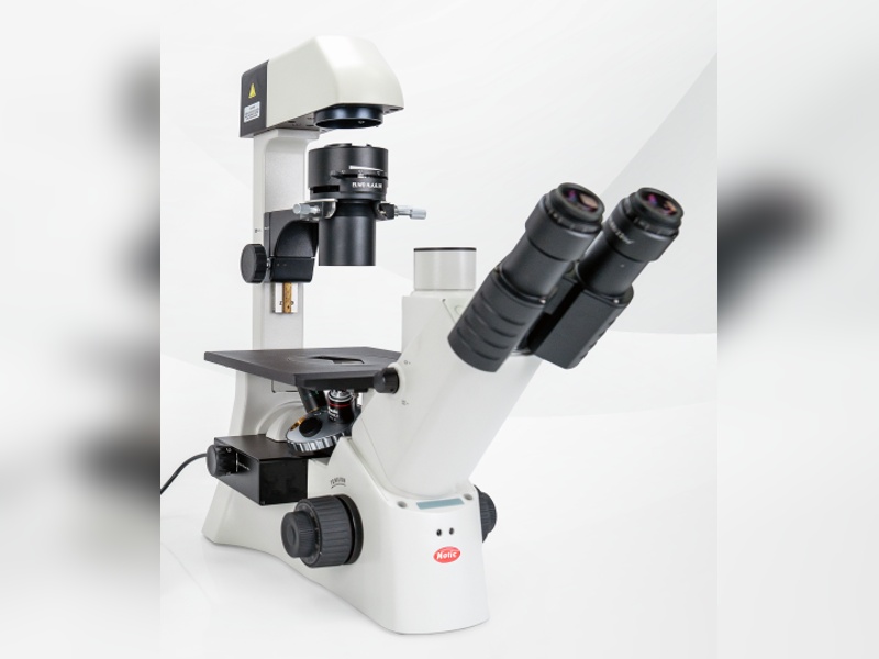
AE31E FL Inverted Fluorescence Microscope
The AE31E FL inverted fluorescence microscope is built for cell cultur
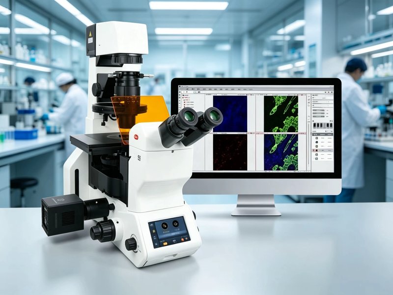
PX55 FS8 Fully Motorized Inverted Fluorescence Microscope
A fully motorized intelligent inverted fluorescence microscope integra
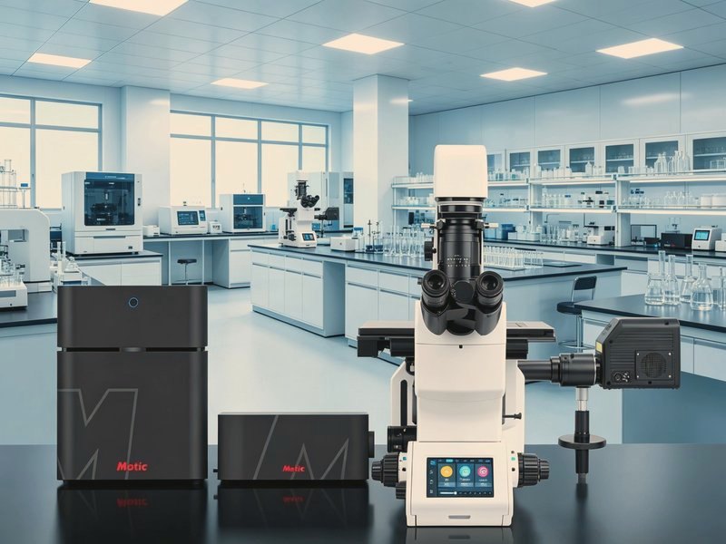
Vertex LS Laser Confocal Microscope
A domestically produced laser scanning confocal imaging and analysis p
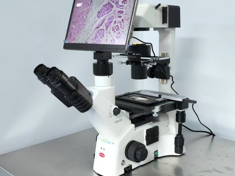
AE31E Inverted Biological Microscope
An inverted biological microscope on the CCIS infinity optical system:
Choosing equipment for this area, or struggling with what you have?
Tell us your sample types, observation goals and daily workload, and we will suggest a configuration stage by stage — no brand decision needed first.