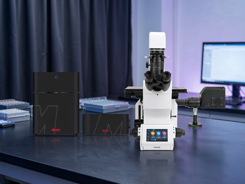
Vertex LS90 Laser Scanning Confocal Microscope
Model: Vertex LS90The Vertex LS90 laser scanning confocal microscope adds a grating-based spectral system with motorised slit to the LS70 platform, resolving 1 nm and separating overlapping fluorescence channels by wavelength. A 25 mm field, 8192 x 8192 scanning and 120 nm XY super-resolution suit plant, pathology and neuroscience samples with strong autofluorescence or overlapping dye spectra.
Specifications
| Spectral scanning and unmixing | Grating dispersion with motorised slit, 1 nm spectral resolution; detector tuning accuracy <= 1 nm |
|---|---|
| Max scan resolution | 8192 x 8192 |
| Scan speed | 8 fps at 512 x 512; up to 30 fps with resonant scanner |
| Super-resolution (deconvolution) | XY 120 nm, Z 300 nm |
| Scan field | 25 mm, 360 degree rotation |
| Bit depth | 16 bit |
| Scan modes | X, Y, Z, lambda, T in any combination |
| Pinhole | Fixed 30 um / motorised continuous / custom fixed |
| Lasers | 405, 488, 561, 640 nm standard at 50 mW each; optional 445, 520, 594, 785 nm |
| Detectors | 4 multi-alkali PMTs, QE >= 28% at 500 nm; GaAsP option, QE >= 45% |
| Transmitted detector | 1 channel, simultaneous brightfield / phase capture with automatic merge |
| Host microscope | PX55 FS8 Plus fully motorised inverted stand |
| Observation modes | Brightfield, phase contrast, fluorescence, DIC, Hoffman, polarisation |
| Eyepiece | 10X / 25 mm ultra-wide field |
| Objective nosepiece | Motorised six-position, coded |
| Condenser | Motorised seven-position long working distance, NA 0.55, WD >= 27 mm |
| Stage | Motorised, 120 x 80 mm travel, repeatability <= 1 um, step 0.05 um |
| Z drive | Motorised, 13 mm travel, 10 nm step |
| Transmitted illumination | 10 W LED |
| Fluorescence illumination | 3 or 7 channel LED (350-700 nm, >= 20000 h) or 130 W metal halide |
| Filter turret | Motorised eight-position, magnetic cubes; DAPI / FITC / TRITC included |
| Objectives | Uplan APO 4X/0.16, 10X/0.45, 20X/0.80, 40X/0.95, 60X/1.42 Oil, 100X/1.45 Oil |
| Software | 2D/3D imaging, large-area stitching, live-cell time lapse, FRAP, deconvolution, colocalisation, AI cell segmentation and tracking |
| Workstation | Windows 11 with GPU accelerator |
| Environment | 10-20 C, humidity <= 60% non-condensing |
Features
- Spectral scanning and unmixing: grating dispersion with motorised slit, 1 nm spectral resolution and <= 1 nm detector tuning, separating overlapping channels by wavelength
- 25 mm scan field with 8192 x 8192 pixels, one large image instead of repeated stitching
- Four lasers at 405/488/561/640 nm, 50 mW each, expandable to 445/520/594/785 nm
- Four multi-alkali PMTs at QE >= 28% at 500 nm, with GaAsP option rated QE >= 45%
- 120 nm XY and 300 nm Z super-resolution through adaptive iterative deconvolution on GPU
- 8 fps at 512 x 512, up to 30 fps with the resonant scanner, enough for dynamic events
- Scan head bolted directly to the stand with no fibre link, preserving photon efficiency and long-term stability
- Fully motorised: six-position nosepiece, seven-position condenser, motorised stage and 10 nm Z step
- Software covers spectral unmixing, deconvolution, 3D rendering, large-area stitching, FRAP, colocalisation, AI segmentation and tracking, with an offline perpetual licence
- Local engineers handle installation, calibration and on-site training, with service response based in Zhengzhou
Applications
- Imaging of strongly autofluorescent samples such as leaves and root systems
- Multicolour immunofluorescence with overlapping dye spectra
- Removal and quantification of autofluorescence in pathology sections
- Multichannel protein expression and colocalisation in neuroscience
- Long-term live-cell time lapse and FRAP kinetics
- Multiparameter fluorescence imaging in drug screening
- Projects that build spectral fingerprints using lambda scanning
- Confocal shared facilities in universities and research institutes
- High-end imaging needs in biopharma companies and CROs
- Fluorescent labelling and structural observation in materials science
Frequently Asked Questions
When is the LS90 required?
When sample autofluorescence cannot be suppressed, when two dyes have broadly overlapping emission, or when you need to record the full emission spectrum for unmixing. The LS70 cannot do these; the LS90 spectral system can.
How is spectral unmixing different from swapping filters?
A filter opens a window in wavelength and cannot cut out overlap. Spectral unmixing captures the whole emission spectrum first, then separates it mathematically against known dye spectra, which genuinely removes bleed-through.
What are the installation requirements?
An anti-vibration table, an ambient temperature of 10-20 C, humidity below 60% non-condensing, and roughly 1.5 m of bench width for the stand plus the laser control box.
Want the full specification sheet?
Leave a number and we will send the specification sheet and a quotation for the Vertex LS90 Laser Scanning Confocal Microscope.