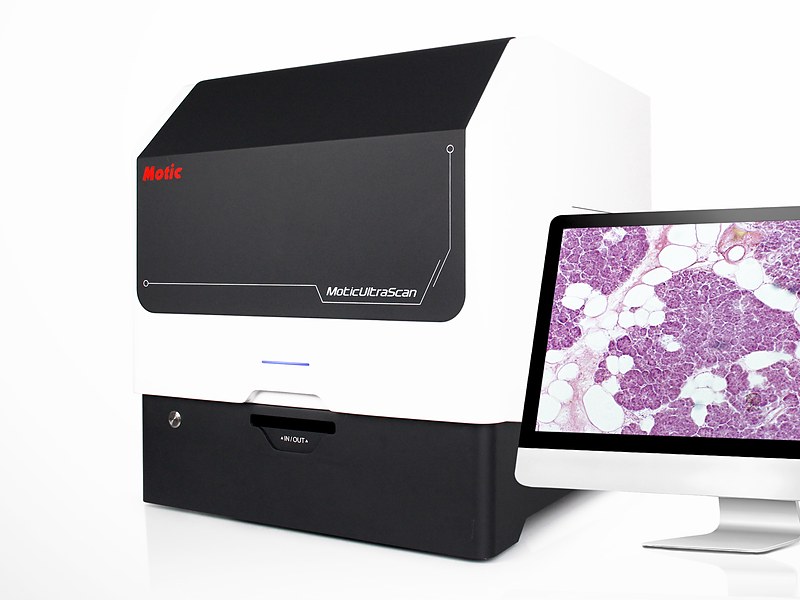
UltraScan 6 Whole Slide Imaging Scanner
Model: UltraScan 6A third-generation fully automated whole slide imaging scanner with a six-slide capacity. A linear magnetic direct-drive stage with grating-scale positioning scans a 15 × 15 mm whole-tissue slide in as little as 20 seconds at 40X, at 0.22 μm per pixel. Five scanning modes — continuous, standard, high-precision, extended depth of field and Z-stack — cover sections from flat to badly uneven, and output in SVS, DICOM, TIFF, CSP and OME-TIFF integrates with remote pathology platforms and AI analysis software.
Specifications
| Slide capacity | 6 slides |
|---|---|
| Objectives | Plan apochromat 20X/0.8 standard; 40X/0.95 and 10X/0.3 optional |
| Objective changer | Motorised, three objectives mounted simultaneously |
| Resolution | 40X mode 0.22 μm/pixel; 20X mode 0.44 μm/pixel; 80X mode 0.11 μm/pixel |
| Scan speed | 15 × 15 mm whole-tissue slide: 25 s at 40X (20 s best case); 70 s at 80X (60 s best case) |
| Scan modes | Continuous, standard, high-precision, extended depth of field, Z-stack |
| Focusing | Real-time autofocus with independent focusing unit |
| Imaging | Three-camera system; 12-megapixel 1.1-inch high-speed primary sensor |
| Illumination | High-frequency stroboscopic LED, synchronised with continuous stage and objective motion |
| Stage | Linear magnetic direct drive with grating-scale positioning, nanometre-level control |
| Slide size | 75–76 × 25–26 mm; optional carrier for double-width 75–76 × 50–51 mm |
| Label reading | Automatic barcode and QR recognition, manual entry and editing supported |
| Image formats | MDS, SVS, JPEG, DICOM, TIFF, OME-TIFF, CSP |
| Colour | ICC colour accreditation supported |
| Quality control | Intelligent per-slide image quality scoring |
| Computer | i7-14700 / 64 GB RAM / 512 GB SSD + 2 TB / RTX 5060 / Windows 11 Professional |
| Interfaces | USB 3.0, 10GigE |
| Standard software | DSAssistant, UltraScanner |
| Optional software | DSServer |
| Dimensions | 585 × 533 × 583 mm |
| Net weight | 51 kg |
Features
- Third-generation scanning platform completes a 15 × 15 mm whole-tissue slide in as little as 20 seconds at 40X, fast enough for intraoperative frozen consultation
- Linear magnetic direct drive with grating-scale positioning delivers nanometre-level accuracy with no drift over long runs
- High-frequency stroboscopic LED illumination captures images while stage and objective move continuously, sparing the slide prolonged intense light
- Five scan modes matched to section flatness: continuous for flat slides, high-precision for sparse ones, extended depth of field for undulating ones, Z-stack for badly uneven ones
- Ultra-flat apochromat objectives keep the whole field flat with colour correction, reproducing the native colour of H&E faithfully
- A 12-megapixel 1.1-inch primary sensor plus a three-camera system balances speed against colour accuracy
- Intelligent per-slide quality scoring stops substandard images from leaving the system
- Motorised changer carries three objectives, and the software recognises carrier formats without manual setup
- Outputs SVS, DICOM, TIFF, CSP and OME-TIFF for integration with laboratory information systems and analysis tools
- Connects to digital pathology remote diagnosis and quality-control platforms and works with any AI pathology software
Applications
- Hospital pathology departments: batch digitisation of routine H&E slides, rapid frozen-section scanning, archiving of difficult cases
- Remote pathology consultation: slides uploaded to the cloud for simultaneous review at referring and specialist hospitals
- Research institutes: histology and pathology research, quantitative morphology, image data archiving
- Medical teaching: building digital slide libraries so students review material on their own computers without timetable or instrument limits
- Pharmaceutical companies and CROs: toxicologic pathology archiving, peer review, multi-centre data sharing
- AI pathology: high-fidelity whole slide images for algorithm training and validation
Frequently Asked Questions
How long does a whole slide scan take at 40X?
A 15 × 15 mm whole-tissue slide scans in about 25 seconds at 40X, with a best case of 20 seconds; at 80X it is about 70 seconds, best case 60. This covers the scan only, excluding sectioning, staining and diagnosis.
Does it integrate with our existing pathology system?
Output supports SVS, DICOM, TIFF, OME-TIFF, CSP and JPEG. DICOM connects directly to PACS and pathology information systems, and other formats are readable by third-party analysis software.
What if the section is folded or uneven?
Match the scan mode to the slide: continuous scanning for flat slides (fastest), high-precision for sparse ones, extended depth of field for undulating ones (multi-layer fusion into one sharp image), and Z-stack for badly uneven sections. Choosing the wrong mode is the main cause of unusable output.
Is a six-slide capacity enough?
At 25 seconds per slide at 40X, six slides take about three minutes. That suits departments running a few dozen slides a day; above 200 per day, consider a higher-capacity model or unattended overnight scanning.
Does scanning damage the slide?
A high-frequency stroboscopic LED fires only at the moment of exposure, so the slide never sits under continuous intense light. That matters for archival slides and fluorescent labels.
Want the full specification sheet?
Leave a number and we will send the specification sheet and a quotation for the UltraScan 6 Whole Slide Imaging Scanner. Calls returned within 10 minutes on working days.