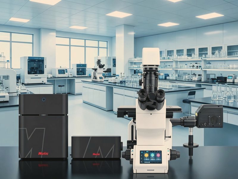
Vertex LS Laser Confocal Microscope
Model: Vertex LSA domestically produced laser scanning confocal imaging and analysis platform with 120nm ultra-high resolution, 8K scanning pixels, 1nm spectral unmixing, and AI intelligent analysis, benchmarked against mid-to-high-end imported systems.
Specifications
| Optical resolution | 120nm ultra-high resolution |
|---|---|
| Imaging speed | Up to 8 fps |
| Scanning pixels | 8192×8192 (8K) |
| Spectral precision | 1nm continuously adjustable, self-developed spectral scanning and unmixing |
| Field of view | 25mm large field of view |
| Detector | Domestically produced PMT high-sensitivity detection |
| Observation mode | Brightfield / Phase contrast / DIC / Fluorescence |
| Stitching | Seamless large-field-of-view stitching |
| AI analysis | Cell quantitative analysis, co-localization analysis, intelligent deconvolution |
| System | Fully domestically produced solution, fully Chinese interface, one-touch startup |
Features
- 120nm ultra-high optical resolution for clear details
- 8192×8192 (8K) scanning pixels for fine imaging
- 8fps high-speed imaging, suitable for dynamic observation
- 1nm adjustable spectral precision for precise unmixing of fluorescence signals
- 25mm large field of view for a large single-acquisition area
- Seamless large-field-of-view stitching for panoramic imaging of large samples
- AI cell quantitative analysis, co-localization analysis, and intelligent deconvolution
- Fully domestically produced solution with fast after-sales response
Applications
- Live dynamic observation and time-series imaging
- 3D reconstruction and Z-axis optical sectioning
- Molecular dynamics experiments such as FRAP
- Cell quantitative analysis and co-localization research
- Biomedical and materials science research
Frequently Asked Questions
How is confocal different from widefield fluorescence?
A pinhole rejects out-of-focus light, so only the focal plane contributes. That enables optical sectioning and 3D reconstruction, whereas widefield sums out-of-focus signal and looks hazier.
What samples suit it?
Multi-channel fluorescence in cells and tissue sections, live-cell dynamics, Z-series of thick samples and 3D reconstruction.
Does it need a dark room?
A darkened environment is recommended. Fluorescence is weak and ambient light noticeably reduces contrast, especially in low-expression samples.
How are laser lines chosen?
By the excitation peaks of the probes: 405, 488, 561 and 640 nm are the common set. Multi-colour work needs attention to channel bleed-through.
Want the full specification sheet?
Leave a number and we will send the specification sheet and a quotation for the Vertex LS Laser Confocal Microscope. Calls returned within 10 minutes on working days.