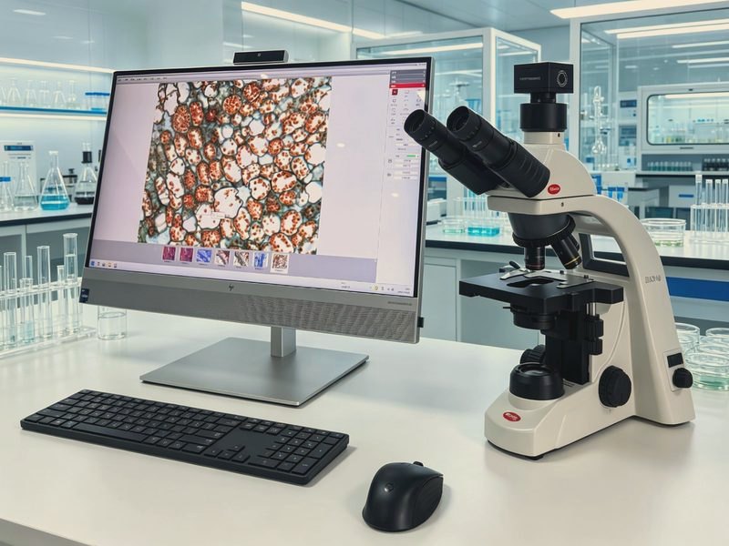
BA310 Digital Imaging Microscope
Model: BA310Based on the high-performance BA310 biological microscope platform, it has a built-in digital imaging port and comes with a same-brand camera and image analysis software, integrating observation, photography, measurement, and analysis.
Specifications
| Optical system | Infinity chromatic aberration-corrected CCIS optical system, multilayer broadband coating (green coating) |
|---|---|
| Eyepiece | 10X/20mm high-eyepoint compensating plan eyepiece, diopter adjustable |
| Objective | Infinity plan achromatic objectives 4X/0.1, 10X/0.25, 20X/0.4, 60X/0.85 (spring) |
| Observation tube | Hinged trinocular, 30° inclined, interpupillary distance 48-75mm, rotatable tube |
| Nosepiece | Inward-tilting 5-hole nosepiece |
| Focusing | Coaxial coarse and fine adjustment, fine 0.2mm/turn, graduation 2μm, with limit stop to prevent slippage |
| Stage | Double-layer compound mechanical moving stage 175×140mm, travel range 76×50mm, with re-positioning function |
| Condenser | N.A.1.25 Abbe condenser with variable aperture diaphragm |
| Illumination | Köhler illumination, 6V3W LED cold light source, lifespan 50,000 hours, continuously adjustable brightness |
| Camera port | Built-in 0.65X camera port |
| Camera | Same-brand sCMOS camera, ≥6.3 million pixels, 1/1.8 inch, USB3.1 |
| Software | Same-brand microscope image analysis software, supporting H&E color correction and automatic segmentation counting |
Features
- CCIS infinity chromatic aberration-corrected optical system with multilayer broadband coating for clear imaging
- EC Plan plan achromatic objectives for high-contrast imaging
- LED cold light source Köhler illumination, lifespan 50,000 hours, continuously adjustable brightness
- Double-layer compound mechanical stage with re-positioning function for precise operation
- Built-in 0.65X camera port for easy expansion to digital imaging
- Same-brand sCMOS camera and microscope image analysis software, compatible and stable
- Supports expansion to phase contrast and darkfield observation, with an overall anti-fungal design
Applications
- Clinical testing and pathology observation
- Biomedical teaching and research
- Observation of cells and tissue sections
- Microscopic analysis of stained specimens
- Digital microscopy teaching and image archiving
Frequently Asked Questions
How many megapixels do I need?
Five to twelve megapixels is enough for routine brightfield work and teaching documentation. For fluorescence and weak signals, pixel size and quantum efficiency matter more than pixel count.
Can fluorescence be added later?
Yes, a reflected-light fluorescence module with LED illumination is available, supporting the usual B, G and U channels for sections and smears.
How should objectives be configured?
Plan achromats at 4X, 10X, 40X and 100X oil are the standard set. For colour photography, use a plan objective at 40X as well so the field edge stays sharp.
What is the difference from an inverted microscope?
On an upright instrument the objective sits above the specimen, which suits slides and smears. On an inverted instrument it sits below, which suits live cells in flasks and dishes.
Want the full specification sheet?
Leave a number and we will send the specification sheet and a quotation for the BA310 Digital Imaging Microscope. Calls returned within 10 minutes on working days.