Confocal versus widefield fluorescence: is it budget or necessity?
A sentence you hear often when buying a fluorescence microscope: start with widefield, move to confocal when it is not enough.
That is half right. Widefield does cover most routine fluorescence work. But what "not enough" actually means is rarely spelled out, so people either overspend or spend years working around a limitation they could have avoided.
1. Where widefield light comes from
A mercury lamp or LED illuminates the whole field, and every excited fluorophore emits at once — above the focal plane as well as below it.
That out-of-focus light enters the objective and lays a fog of background over the image. With thin sections and single layers of cells the effect is mild; with anything thicker, the background swamps the detail.
2. What confocal changes
Confocal places a pinhole in front of the detector, optically conjugate to the objective's focal point.
Light from the focal plane passes; light from elsewhere is blocked. That is optical sectioning — isolating one plane without cutting the sample.
Three practical gains follow.
First, thick samples become readable. Organoids, embryos and thick tissue sections look like mush in widefield; confocal steps through them plane by plane and reconstructs the 3D structure.
Second, resolution improves. The pinhole clips the edge of the focal spot, taking lateral resolution into the 120 nm class — close to double that of widefield.
Third, spectral unmixing becomes possible. When channels overlap, spectral scanning separates the signals, something widefield cannot do.
3. When widefield is genuinely enough
The criterion is not how expensive the sample is, but whether what you need to see lies in one plane.
Routine pathology: H&E, special stains, IHC, with sections 4 to 5 microns thick, essentially one plane;
Monolayer cultures: morphology, fluorescence intensity, transfection efficiency, drug response;
FISH dot counting;
Teaching and routine screening.
Widefield handles all of this, and does it faster, with lower phototoxicity and simpler operation. Confocal's intense light can kill live cells — a reverse cost that is often overlooked.
4. When confocal is unavoidable
When the sample has thickness, and the thickness carries information.
3D architecture of organoids and spheroids;
Neural and vascular networks inside sections tens to hundreds of microns thick;
3D migration, endocytosis and division in live cells;
Co-localisation analysis — proving two proteins really sit at the same place requires rejecting out-of-focus signal;
Sub-cellular structures needing super-resolution.
One more case is easily missed: quantitation of low-abundance signal. Widefield background lifts the baseline and buries weak signal; confocal suppresses the background so the weak signal surfaces.
5. Which numbers to specify
Spectral precision. Channel overlap is normal in multicolour work; 1 nm continuously tunable scanning is what separates DAPI, FITC, TRITC and tissue autofluorescence.
Scan pixels and field of view. An 8K scan with a 25 mm field covers a large area per acquisition and leaves fewer seams in large-area stitching.
Speed. Live-cell work wants seconds or faster. 8 fps is not fast for large frames, but it is enough for dynamic processes.
Detector. Sensitivity determines whether weak signal is captured at all, and whether laser power can be turned down to spare the sample.
Software. Deconvolution, co-localisation analysis and cell counting save post-processing time later.
6. Two misconceptions
First, that confocal simply means sharper fluorescence. It is sharper not because the optics are better, but because out-of-focus light is removed. If the sample is already thin, the gain is limited and the money is poorly spent.
Second, that confocal requires the fullest possible laser set. Lasers should follow the excitation peaks of your probes. An extra unusable wavelength is an idle instrument you paid for.
Summary
Widefield and confocal are not substitutes but a division of labour. Routine reading, monolayers and teaching screening belong to widefield; 3D structure, thick samples, co-localisation and weak signal belong to confocal. The real question is not which one to buy, but whether your samples have thickness.
Equipment mentioned
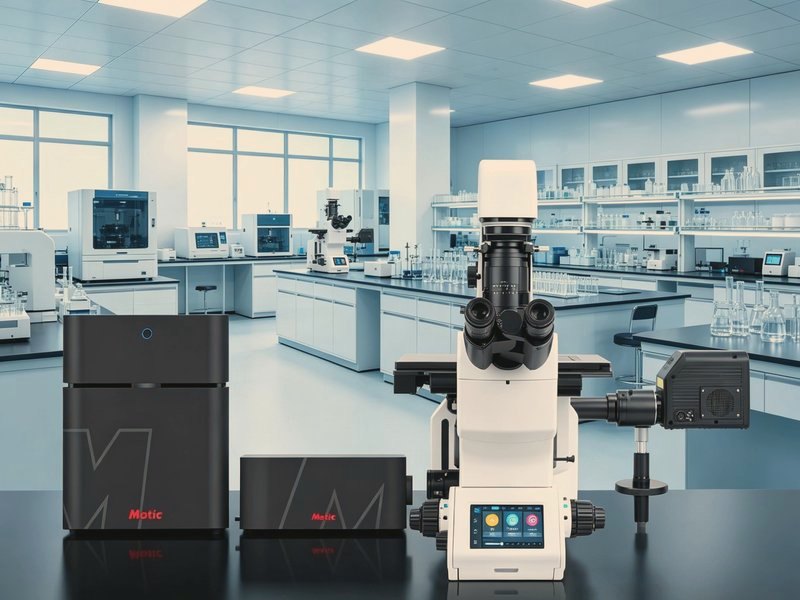
Vertex LS Laser Confocal Microscope
A domestically produced laser scanning confocal imaging and
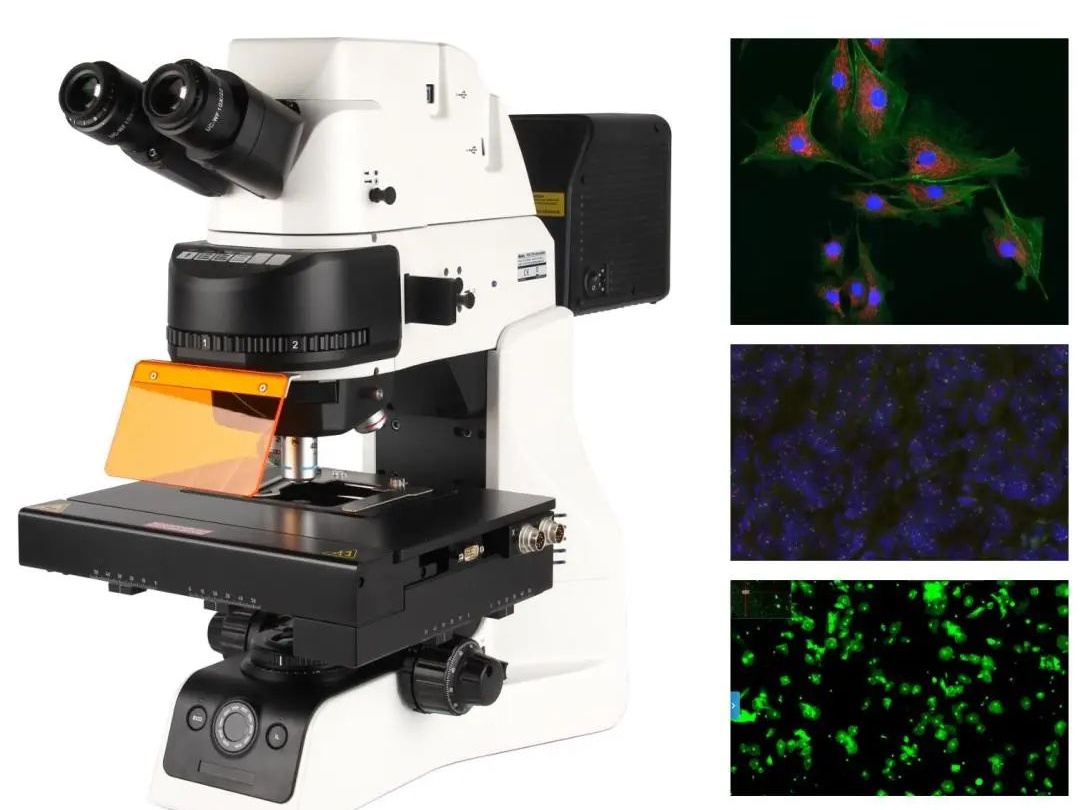
PA53 FS6 Inverted Fluorescence Microscope
The PA53 FS6 is a research-grade upright fluorescence micros
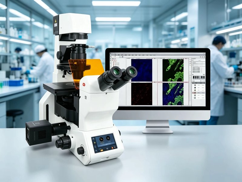
PX55 FS8 Fully Motorized Inverted Fluorescence Microscope
A fully motorized intelligent inverted fluorescence microsco