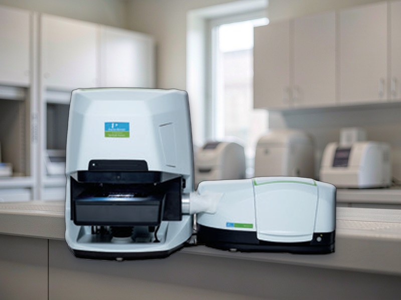
Microplastics, Coating Defects, Tablet Contaminants: How an IR Microscope Finds Invisible Chemical Differences
An optical microscope answers where, how big and what shape. But a 20 µm black particle could be carbon black, iron oxide, or a degraded polymer fragment. The image alone cannot tell.
The FT-IR microscope works differently: locate the target in visible light, pass infrared light through that exact spot, collect its absorption spectrum and match it against a library. One instrument, both a microscope and an identification tool.
1. Spatial resolution: what 7 µm buys you
Infrared wavelengths are an order of magnitude longer than visible light, so diffraction limits IR microscopy to a few to a dozen micrometres.
Better than 7 µm means particles above roughly 10 µm can be identified point by point without pre-concentration. Smaller particles require line or area mapping, combining spectra from several pixels.
Reports should always state the smallest identifiable particle size and the detection limit, otherwise data from different labs cannot be compared.
2. Three sampling modes
Transmission suits IR-transparent samples: thin films, particles on filters, microtome sections. Fast, but thick samples absorb too strongly.
Reflection suits opaque surfaces such as contaminants on metal or coating cross-sections.
Micro-ATR is the most universal. A diamond or germanium tip is pressed onto the sample with no preparation, handling bulk pieces, fibres and thick sections. Magnetic tips swap easily, but contact pressure must adapt automatically — too much pressure leaves marks on soft samples and distorts the result.
3. Curved and rough samples: Z-STACK fixes focus
Classical IR microscopy assumes a flat sample; height differences beyond the depth of field ruin the measurement.
Z-STACK focuses layer by layer along the Z axis, combines the sharp regions into one fully focused image and records depth information for 3D reconstruction.
In practice this means circuit boards, oddly shaped components and rough surfaces go straight onto the stage — no polishing, no embedding. For failure analysis and foreign matter investigations, that saves both time and the risk of destroying the sample.
4. Why microplastics need it
Microplastic identification relies on infrared or Raman spectra; morphology alone cannot determine polymer type. The accepted criterion is a library match score above a threshold, commonly 70%.
Automation is where the value lies: scan the whole filter, detect particles, acquire spectra one by one, count by polymer type and report size distribution.
A 47 mm filter that takes a full day to inspect manually yields a complete report in a few hours, with consistent, reproducible counting criteria.
5. Deciding whether a system is adequate
Three things: smallest identifiable particle size, maximum sample thickness, and degree of automation.
The first two decide whether it can see and hold the sample; the third decides whether it can actually do the work. Put those three into tender specifications — they matter far more than a list of optical components.
Summary
The value of an IR microscope is not its resolution figure but the chain it builds between image and composition. The shorter that chain, the faster a finding turns into a conclusion.