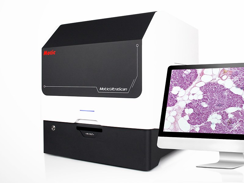
UltraScan 6 Whole Slide Imaging Scanner
Model: UltraScan 6A third-generation fully automated whole slide imaging scanner with a six-slide capacity. A linear magnetic direct-drive stage with grating-scale positioning scans a 15 × 15 mm whole-tissue slide in as little as 20 seconds at 40X, at 0.22 μm per pixel. Five scanning modes — continuous, standard, high-precision, extended depth of field and Z-stack — cover sections from flat to badly uneven, and output in SVS, DICOM, TIFF, CSP and OME-TIFF integrates with remote pathology platforms and AI analysis software.
Specifications
| Slide capacity | 6 slides |
|---|---|
| Objectives | Plan apochromat 20X/0.8 standard; 40X/0.95 and 10X/0.3 optional |
| Objective changer | Motorised, three objectives mounted simultaneously |
| Resolution | 40X mode 0.22 μm/pixel; 20X mode 0.44 μm/pixel; 80X mode 0.11 μm/pixel |
| Scan speed | 15 × 15 mm whole-tissue slide: 25 s at 40X (20 s best case); 70 s at 80X (60 s best case) |
| Scan modes | Continuous, standard, high-precision, extended depth of field, Z-stack |
| Focusing | Real-time autofocus with independent focusing unit |
| Imaging | Three-camera system; 12-megapixel 1.1-inch high-speed primary sensor |
| Illumination | High-frequency stroboscopic LED, synchronised with continuous stage and objective motion |
| Stage | Linear magnetic direct drive with grating-scale positioning, nanometre-level control |
| Slide size | 75–76 × 25–26 mm; optional carrier for double-width 75–76 × 50–51 mm |
| Label reading | Automatic barcode and QR recognition, manual entry and editing supported |
| Image formats | MDS, SVS, JPEG, DICOM, TIFF, OME-TIFF, CSP |
| Colour | ICC colour accreditation supported |
| Quality control | Intelligent per-slide image quality scoring |
| Computer | i7-14700 / 64 GB RAM / 512 GB SSD + 2 TB / RTX 5060 / Windows 11 Professional |
| Interfaces | USB 3.0, 10GigE |
| Standard software | DSAssistant, UltraScanner |
| Optional software | DSServer |
| Dimensions | 585 × 533 × 583 mm |
| Net weight | 51 kg |
Features
- Third-generation scanning platform completes a 15 × 15 mm whole-tissue slide in as little as 20 seconds at 40X, fast enough for intraoperative frozen consultation
- Linear magnetic direct drive with grating-scale positioning delivers nanometre-level accuracy with no drift over long runs
- High-frequency stroboscopic LED illumination captures images while stage and objective move continuously, sparing the slide prolonged intense light
- Five scan modes matched to section flatness: continuous for flat slides, high-precision for sparse ones, extended depth of field for undulating ones, Z-stack for badly uneven ones
- Ultra-flat apochromat objectives keep the whole field flat with colour correction, reproducing the native colour of H&E faithfully
- A 12-megapixel 1.1-inch primary sensor plus a three-camera system balances speed against colour accuracy
- Intelligent per-slide quality scoring stops substandard images from leaving the system
- Motorised changer carries three objectives, and the software recognises carrier formats without manual setup
- Outputs SVS, DICOM, TIFF, CSP and OME-TIFF for integration with laboratory information systems and analysis tools
- Connects to digital pathology remote diagnosis and quality-control platforms and works with any AI pathology software
Applications
- Hospital pathology departments: batch digitisation of routine H&E slides, rapid frozen-section scanning, archiving of difficult cases
- Remote pathology consultation: slides uploaded to the cloud for simultaneous review at referring and specialist hospitals
- Research institutes: histology and pathology research, quantitative morphology, image data archiving
- Medical teaching: building digital slide libraries so students review material on their own computers without timetable or instrument limits
- Pharmaceutical companies and CROs: toxicologic pathology archiving, peer review, multi-centre data sharing
- AI pathology: high-fidelity whole slide images for algorithm training and validation