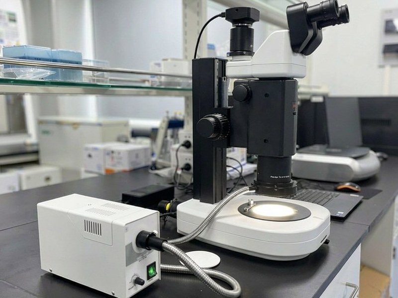
SM20 Research-Grade Stereo Microscope
Model: SM20A research-grade stereo microscope with a 20:1 zoom ratio, covering everything from a low-power overview to high-power detail on a single instrument — for life science research and precision industrial inspection.
Specifications
| Optical system | Parallel (CMO, Galilean) optical system |
|---|---|
| Head | Trinocular, visual/photo split 100:0 / 80:20 |
| Interpupillary | 55-75mm; one tube adjusts 180° vertically, 130mm viewing height range |
| Zoom ratio | 20:1 |
| Zoom range | 0.75X-15X (7.5X-150X total with standard 1X plan apochromat) |
| Detents | 11 positions: 0.75, 1, 2, 3, 4, 5, 6, 8, 10, 12, 13.5, 15 |
| Eyepiece | 10X/25 with ±5 dioptre adjustment (standard); 15X/16, 20X/13 (optional) |
| Objectives | Plan apochromat 0.5X N.A.0.075 WD71mm; 1X N.A.0.16 WD60mm; 1.6X N.A.0.24 WD30mm; 2X N.A.0.32 WD20mm |
| Objective mount | M61×1 thread |
| Max field of view | 33.3mm |
| Max resolution | 900 lp/mm |
| Max depth of field | 2.8mm |
| Stand | Upright transmitted-LED base |
| Focusing | Coaxial coarse/fine, adjustable tension, 120mm travel |
| Illumination | Transmitted LED with oblique contrast adjustment and aperture switching; 10W ring reflected LED; 50,000+ hours |
| Optional | Darkfield illuminator, single and combined polarisers, ring fibre illuminator, gem clip, K-series sliding stage (φ95mm), X-Y moving stage (φ95mm) |
Features
- 20:1 continuous zoom — from low-power overview to high-power detail without changing objectives
- Plan apochromat objectives: low distortion, longer working distance
- Galilean optics for flat field and faithful colour reproduction
- One tube adjusts 180° vertically, giving a 130mm viewing-height range for different users
- 25mm wide-field eyepieces, maximum field of view over 33mm
- Coaxial coarse/fine focus with 120mm travel
- Transmitted LED with oblique contrast control for transparent and semi-transparent samples
- 10W ring reflected LED for bright, even, high-contrast illumination
- Expandable with darkfield, polarising and fluorescence accessories; supports Moticam imaging
Applications
- Live observation of model organisms (Drosophila, C. elegans, zebrafish)
- Embryology and developmental biology
- Insect dissection and morphology
- Precision parts and 3D structure inspection
- High-reflectance surface examination
- Jewellery, textiles and forensic evidence