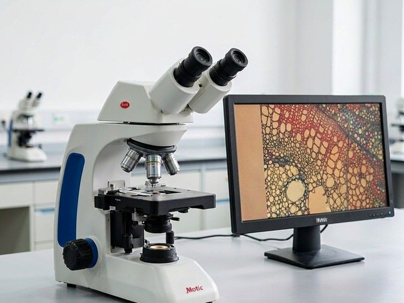
M200D Biological Digital Microscope
Model: M200DA teaching-grade biological microscope with fully integrated digital imaging: an 8-megapixel camera is built into the head, on an infinity-corrected optical system, for classroom demonstration and routine biological observation.
Specifications
| Optical system | Infinity-corrected independent chromatic aberration correction, with built-in digital imaging module |
|---|---|
| Head | Hinged binocular, 30° inclined, interpupillary distance 55-75mm |
| Eyepiece | Wide field 10X/20mm, dioptre adjustable on both tubes, broadband coating with ~95% transmission |
| Objectives | Broadband coated plan achromat 4X / 10X / 40X (spring) / 100X (spring, oil) |
| Objective specs | 4X N.A.0.1 WD≥15.5mm; 10X N.A.0.25 WD≥7mm; 40X N.A.0.65 WD≥0.71mm; 100X N.A.1.25 WD≥0.14mm |
| Nosepiece | Inward-inclined quadruple nosepiece |
| Condenser | Abbe N.A.1.25, accepts simple darkfield, phase contrast and polarising accessories |
| Stage | Arc design, ≥140×140mm, travel ≥75×50mm, vertical travel 22mm, hard anodised surface |
| Slide holder | Reverse-chamfer design, prevents slides lifting during 100X oil observation |
| Focusing | Coaxial coarse and fine, with upper limit stop, fine 0.2mm/turn, graduation 0.002mm |
| Illumination | 3W LED, approx. 60,000 hours lifetime |
| Built-in camera | ≥8 megapixel colour sensor, USB 3.0 output |
| Software | Images 3.0 microscopy image analysis suite |
| Power | Single external DC supply, front I/O switch separated from light control |
| Cooling | 3.5mm gap air-induction cooling in the base |
| Portability | Rear carry handle |
Features
- Camera fully integrated into the microscope head, no extra footprint
- 8 megapixel colour sensor with USB 3.0 output
- Infinity-corrected optical system for high image contrast
- Broadband coated plan achromat objectives, flat across the field
- 3W LED illumination with approx. 60,000-hour lifetime
- Images 3.0 software with side-by-side comparison, cell counting and optical density statistics
- Colour correction mode for H&E stained specimens
- LAN image sharing so other terminals can view the live image
- Single external DC supply with front switch separated from brightness control
Applications
- Classroom demonstration and multimedia teaching
- Routine biological section observation
- Cell morphology and cell counting
- Microbial smear examination
- Preliminary pathology observation
- Everyday laboratory documentation