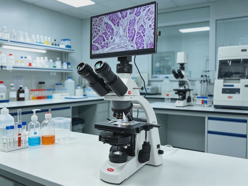
BA310 Tablet Digital Microscope
Model: BA310Based on the BA310 biological microscope platform, it integrates digital imaging with an all-in-one tablet display, enabling real-time observation, photography, and teaching demonstrations without an external computer.
Specifications
| Optical system | Infinity chromatic aberration-corrected CCIS optical system, multilayer broadband coating (green coating) |
|---|---|
| Eyepiece | 10X/20mm high-eyepoint compensating plan eyepiece, diopter adjustable |
| Objective | Infinity plan achromatic objectives 4X/0.1, 10X/0.25, 20X/0.4, 60X/0.85 (spring) |
| Observation tube | Hinged trinocular, 30° inclined, interpupillary distance 48-75mm |
| Nosepiece | Inward-tilting 5-hole nosepiece |
| Stage | Double-layer compound mechanical moving stage 175×140mm, travel range 76×50mm |
| Condenser | N.A.1.25 Abbe condenser with variable aperture diaphragm |
| Illumination | Köhler illumination, 6V3W LED cold light source, lifespan 50,000 hours, continuously adjustable brightness |
| Display | All-in-one tablet display with real-time screen mirroring, no external computer required |
| Camera | Same-brand digital camera, HD imaging |
| Software | Same-brand microscope image analysis software, supporting photography, measurement, and annotation |
Features
- All-in-one tablet display with real-time screen mirroring, no external computer required
- CCIS infinity chromatic aberration-corrected optical system with multilayer broadband coating
- EC Plan plan achromatic objectives for high-contrast imaging
- LED cold light source Köhler illumination, lifespan 50,000 hours, continuously adjustable brightness
- Double-layer compound mechanical stage with re-positioning function
- Same-brand digital camera and image analysis software, compatible and stable
- Suitable for teaching demonstrations and simultaneous viewing by multiple people
Applications
- Teaching demonstrations and simultaneous viewing by multiple people
- Clinical testing and pathology observation
- Biomedical teaching and research
- Observation of cells and tissue sections
- Digital microscopy teaching and image archiving