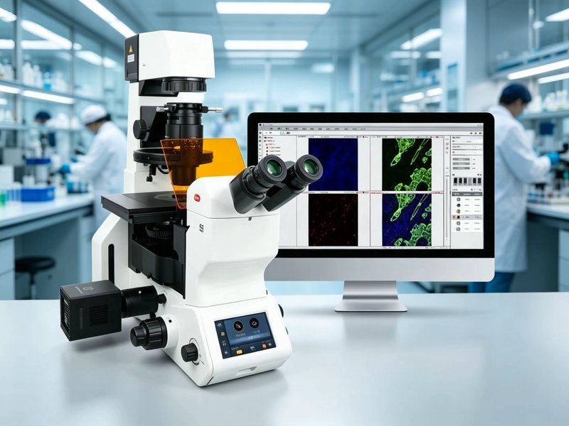
PX55 FS8 Fully Motorized Inverted Fluorescence Microscope
Model: PX55 FS8A fully motorized intelligent inverted fluorescence microscope integrating a CCIS infinity optical system, a motorized coded nosepiece, and an intelligent touchscreen, suitable for cell microscopy imaging and research across multiple fields.
Specifications
| Optical system | CCIS infinity corrected optical system |
|---|---|
| Observation mode | Brightfield / Phase contrast / Fluorescence / Hoffman / DIC / Polarized light / Darkfield |
| Fluorescence light source | Supports LUMOS3 LED fluorescence lamp, lifespan over 20,000 hours, ready to use with low phototoxicity |
| Fluorescence filter blocks | Standard DAPI/FITC/TRITC, 7+1 channel intelligent coded turret |
| Nosepiece | Motorized coded 6-hole nosepiece, automatically recognizing objective magnification |
| Objective | Plan Fluor LWD 10X/20X/40X fluorescence phase contrast, LD Plan Fluor 60X |
| Stage | XY motorized hard-coated stage, travel 130×90mm |
| Condenser | Long working distance, N.A.0.3/0.55, working distance 72mm/27mm |
| Focusing | Coaxial coarse and fine adjustment, coarse 42mm/turn, fine 0.1mm/turn |
| Observation tube | Hinged 45° trinocular, 360° rotation, adjustable beam split ratio |
| Touchscreen | Front-mounted 5-inch OLED touchscreen, displaying device status in real time |
| Camera | Same-brand global shutter camera, 2/3 inch, 2448×2048, USB3.1 |
| Software | Same-brand research and analysis software, supporting real-time EDF, stitching, automatic counting, and multichannel fluorescence |
Features
- Fully motorized control: motorized nosepiece, motorized stage, and motorized filter block turret
- Intelligent coded recognition automatically displaying the current objective magnification
- 7+1 channel intelligent coded fluorescence turret with automatic position recognition
- LUMOS3 LED fluorescence light source, lifespan over 20,000 hours, low phototoxicity, ready to use
- Front-mounted 5-inch OLED touchscreen displaying device status in real time
- Supports multiple observation modes such as brightfield, phase contrast, fluorescence, and DIC
- Same-brand global shutter camera and research and analysis software with powerful functions
Applications
- Cell microscopy imaging and live cell observation
- Immunofluorescence and multi-label imaging
- Histology and pathology observation
- Microbiology and medical research
- Pharmaceutical R&D and biotechnology