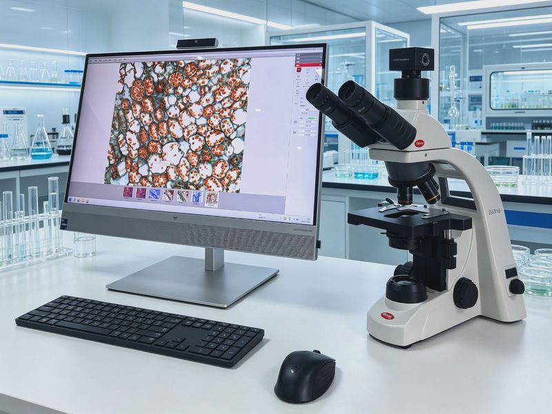
BA310 Digital Imaging Microscope
Model: BA310Based on the high-performance BA310 biological microscope platform, it has a built-in digital imaging port and comes with a same-brand camera and image analysis software, integrating observation, photography, measurement, and analysis.
Specifications
| Optical system | Infinity chromatic aberration-corrected CCIS optical system, multilayer broadband coating (green coating) |
|---|---|
| Eyepiece | 10X/20mm high-eyepoint compensating plan eyepiece, diopter adjustable |
| Objective | Infinity plan achromatic objectives 4X/0.1, 10X/0.25, 20X/0.4, 60X/0.85 (spring) |
| Observation tube | Hinged trinocular, 30° inclined, interpupillary distance 48-75mm, rotatable tube |
| Nosepiece | Inward-tilting 5-hole nosepiece |
| Focusing | Coaxial coarse and fine adjustment, fine 0.2mm/turn, graduation 2μm, with limit stop to prevent slippage |
| Stage | Double-layer compound mechanical moving stage 175×140mm, travel range 76×50mm, with re-positioning function |
| Condenser | N.A.1.25 Abbe condenser with variable aperture diaphragm |
| Illumination | Köhler illumination, 6V3W LED cold light source, lifespan 50,000 hours, continuously adjustable brightness |
| Camera port | Built-in 0.65X camera port |
| Camera | Same-brand sCMOS camera, ≥6.3 million pixels, 1/1.8 inch, USB3.1 |
| Software | Same-brand microscope image analysis software, supporting H&E color correction and automatic segmentation counting |
Features
- CCIS infinity chromatic aberration-corrected optical system with multilayer broadband coating for clear imaging
- EC Plan plan achromatic objectives for high-contrast imaging
- LED cold light source Köhler illumination, lifespan 50,000 hours, continuously adjustable brightness
- Double-layer compound mechanical stage with re-positioning function for precise operation
- Built-in 0.65X camera port for easy expansion to digital imaging
- Same-brand sCMOS camera and microscope image analysis software, compatible and stable
- Supports expansion to phase contrast and darkfield observation, with an overall anti-fungal design
Applications
- Clinical testing and pathology observation
- Biomedical teaching and research
- Observation of cells and tissue sections
- Microscopic analysis of stained specimens
- Digital microscopy teaching and image archiving