Project Overview
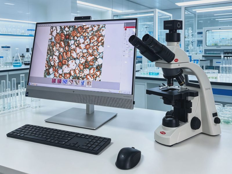
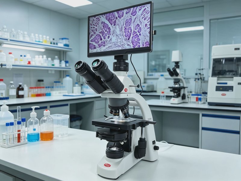
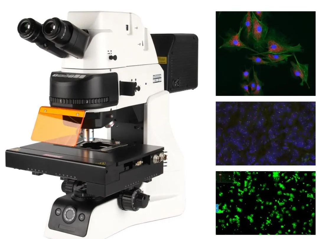
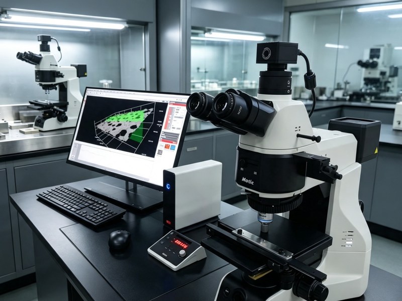
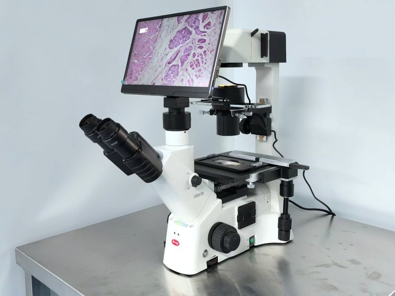
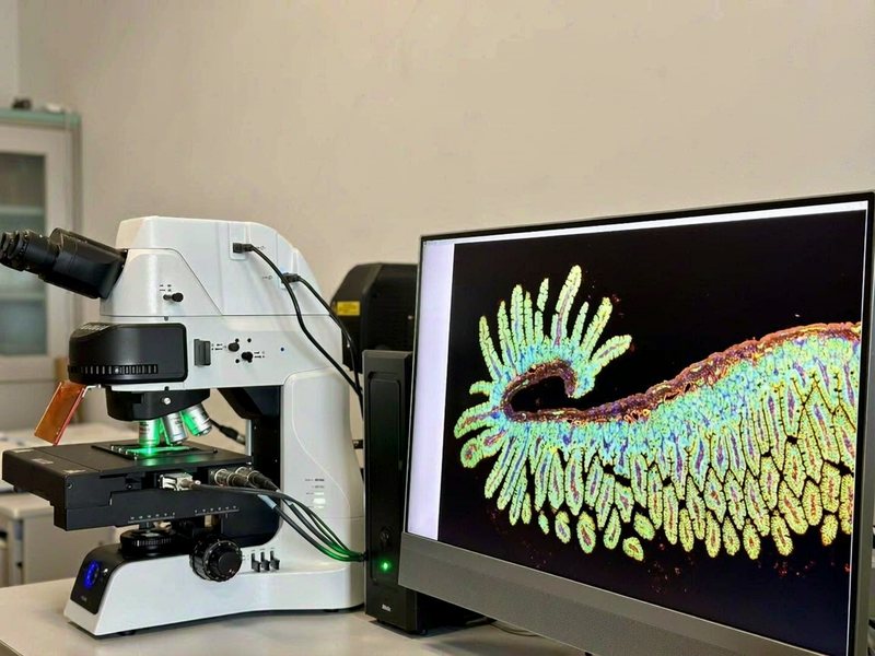
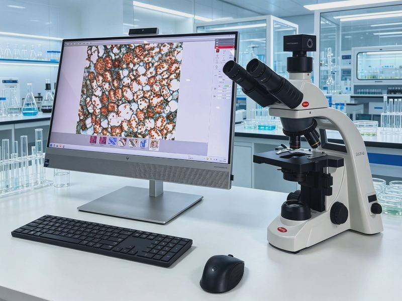
Delivered a BA310 D digital biological microscope with digital imaging to the centre, for colony observation and Gram-stain microscopy in food microbiology, longitudinal and cross-sectional fibre identification in textiles, and image capture and archiving.
Why would a retailer build its own testing centre? The answer sits on the receiving bench every day: goods move from supplier to shelf, and three things have to happen in between — incoming acceptance, routine sampling and traceability when something goes wrong. Fresh produce, bakery and prepared foods need microbiological checks; textiles need fibre composition; and when a problem appears, evidence has to be produced.
1. Two sample types, two observation routes
Food microbiology runs from macro to micro: first the colonies on the plate, then stained slides under the microscope.
Textile fibres are identified by morphology: longitudinal and cross-sectional appearance tell you what the fibre is.
The magnifications, illumination and imaging priorities differ, yet both usually land on the same bench, used by the same team in rotation.
2. Food microbiology: from colony to cell
Total plate count, coliforms, moulds and yeasts all start with colony morphology — size, edge regularity, colour, elevation, smooth or wrinkled surface. This is low-power work where a flat field matters: if edges blur, the same colony gets recorded as two different morphologies, and counting becomes unreliable.
Suspicious colonies then go to Gram staining. Under oil immersion the cell shape and staining reaction decide: Gram-positive purple, Gram-negative red; cocci or bacilli, chains or clusters. This step is the most demanding on light path cleanliness and colour fidelity — a colour shift in the stain leads directly to a wrong reading.
Mould identification goes one level deeper. Aspergillus vesicle shape, Penicillium broom-like conidiophores, Mucor sporangia — these structures only separate at medium to high power, and they depend on contrast and depth of field.
3. Textile fibres: morphology is the evidence
Fibre identification does not rely on reagents, it relies on shape.
Cotton shows natural convolutions, like a flattened ribbon; hemp and flax carry cross-markings and longitudinal striations; wool is covered in scales with serrated edges; standard polyester and nylon appear relatively smooth; regenerated fibres usually show serrated or irregular circular cross-sections.
Preliminary blend ratio estimates, and confirmation that a "100% cotton" label contains synthetic fibre, both rest on these morphological differences. The requirements are concrete: enough contrast, sharp edges, and the ability to capture the typical morphology into a report — "it looked like polyester" will not survive review.
4. What the BA310 D contributes to both jobs
Plan objectives come first. Sharp from centre to edge means no hunting for focus while scanning colonies, and a whole fibre visible in one view.
Integrated digital imaging solves the old external-camera problem. Camera and body share one light path, so observing and capturing no longer compete; a single USB connection to a PC gives live display and acquisition without extra hardware or adapters.
The bundled imaging software covers the reporting step: measuring cell length and fibre diameter, adding scale bars and annotations, exporting images in batches. The record becomes image plus data rather than a sentence of description.
LED illumination keeps things stable. Colour temperature does not drift with running time, so slides imaged on the early and late shift match, and reports remain comparable.
5. Delivery and training
On arrival the system was optically aligned, with camera white balance and exposure parameters calibrated. Training ran in two rounds: daily observation and capture, then measurement, annotation and report output.
Five fixed scenarios — colonies, Gram stains, moulds, fibre longitudinal view, fibre cross-section — were documented as templates with their objective magnification, light level and exposure settings. When staff change, a newcomer following the template produces images that line up with earlier ones.
6. Outcome
The instrument now covers the centre's routine food microbiology microscopy and textile fibre identification, with captured images archived alongside the inspection records.
For a retailer's testing centre, a microscope is not research equipment — it is a tool that produces conclusions every day. Seeing clearly, capturing reliably and archiving properly matter more than magnification figures on a datasheet.