The Pathology Microscope: Four Different Jobs in One Day
How often a pathology department actually uses its microscopes
From outside, pathology looks like a place where slides are read. From inside, the microscope barely stops: gross examination during sampling, slides after staining, difficult cases at review, intraoperative frozen sections that must reach a conclusion in minutes, and teaching or consultation where the field has to be shown to someone else.
One instrument carries several jobs, so selection cannot rest on "can it see clearly" alone.
1. Four stages, four different requirements
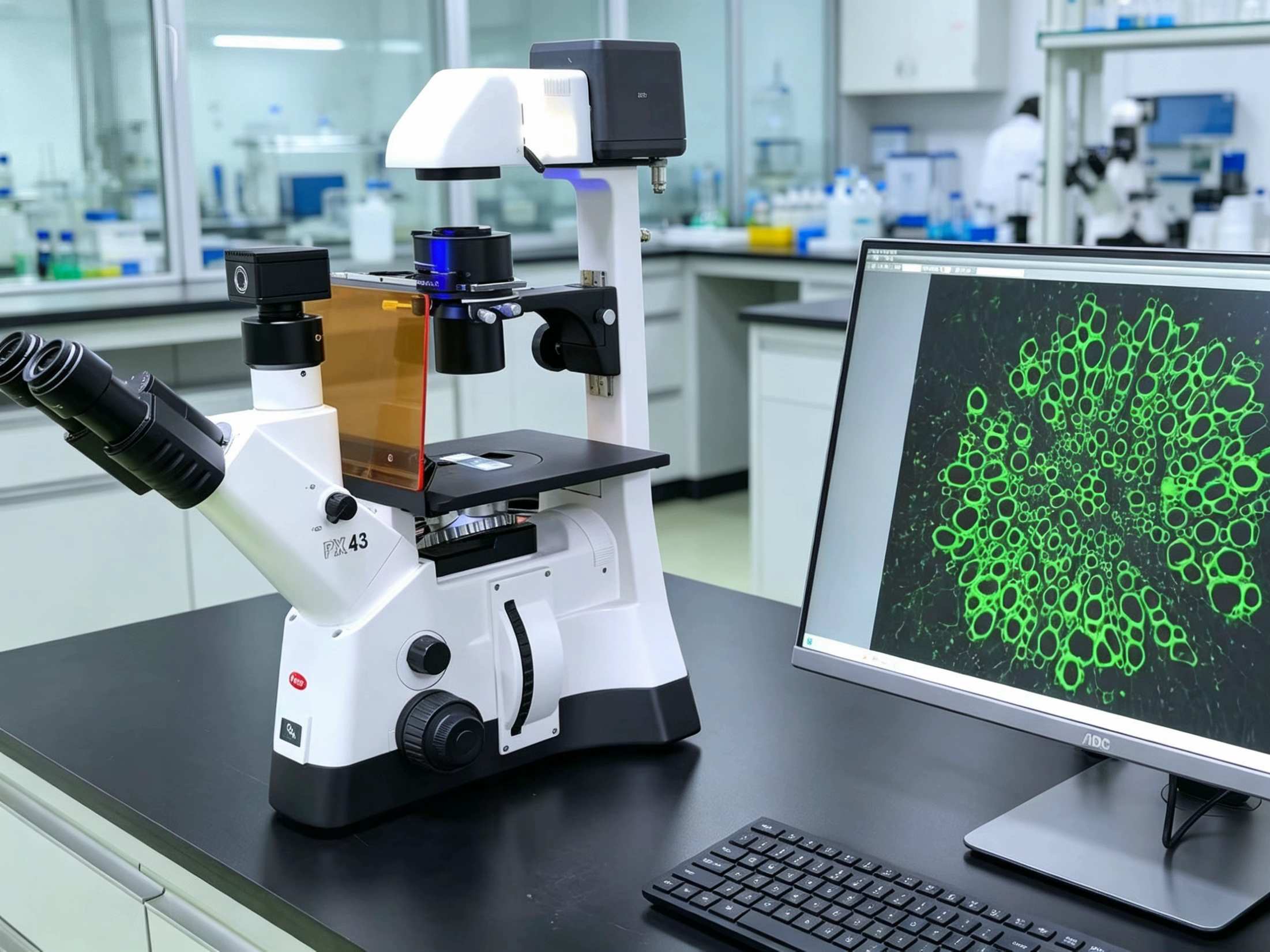
Screening and routine diagnosis. The requirement is comfort and stability over long sessions. A wide field of view, smooth focusing and no eye strain after hours of seated work. No elaborate features are needed, but mechanical precision must be good.
Intraoperative frozen section. The requirement is speed. Slides must be read immediately, often for margin assessment, with ample light and clear contrast. This stage is more sensitive to responsiveness than to resolution.
Difficult cases and teaching. The requirement is two people viewing simultaneously. A dual or multi-head eyepiece lets two pathologists see the same field, which changes the efficiency of discussion entirely. This configuration is undervalued in many departments.
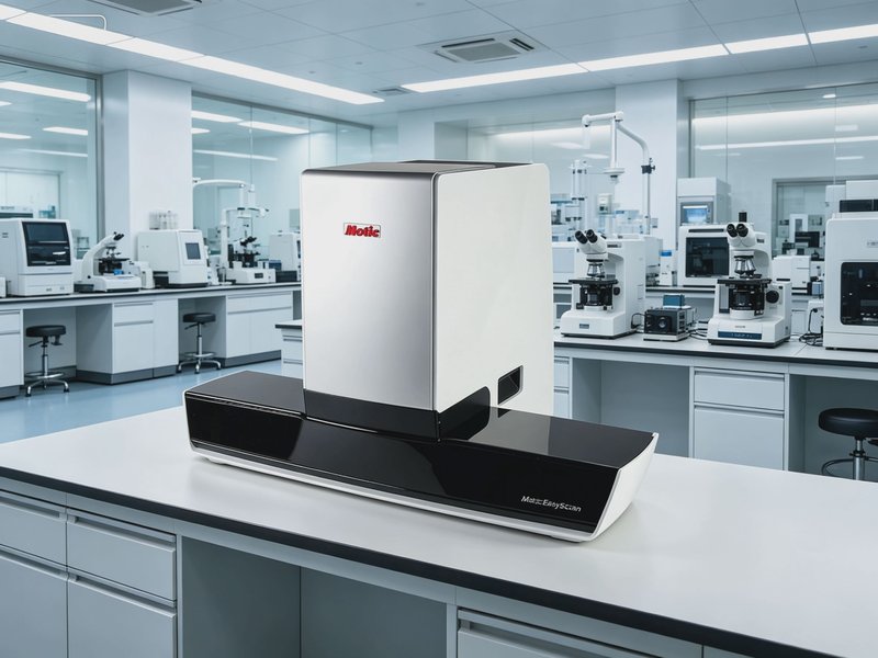
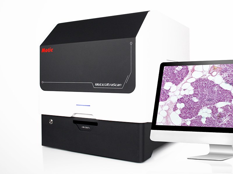
Consultation and remote review. The requirement is to send the field elsewhere. A digital slide scanner turns a whole slide into a zoomable image, which serves remote consultation, teaching and archiving.
2. How far to go with digitisation
Most tertiary hospital departments now have a slide scanner, but the uses vary widely.
High-value uses: remote consultation, departmental archiving, trainee self-study, teaching. Here the scanner earns its place immediately.
Low-value uses: routine screening. Most departments still read directly down the microscope; the scanner supplements rather than replaces.
The test is practical. If consultations and teaching together amount to a handful per month, equip the microscopes and dual eyepieces first and defer the scanner.
3. Two things easily overlooked
Staining quality sets the ceiling. On the same microscope, good staining and poor staining look like two different instruments. Judge equipment without judging the slides and you will attribute a staining problem to the optics.
Calibration and maintenance. Diagnostic pathology does not measure dimensions, but equipment still needs periodic maintenance: cleaning the optical path, checking light-source colour temperature, verifying brightness consistency. Whether this is done determines whether two pathologists reach the same conclusion on the same slide.
Closing note
The logic for a pathology department is tiered configuration by stage, not one instrument that does everything. Screening needs comfort, frozen section needs speed, teaching needs two viewers, consultation needs to transmit.
If your department is renewing equipment, tell us the daily slide volume, consultation frequency and teaching load, and we will advise stage by stage.