Project Overview
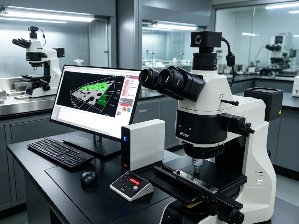
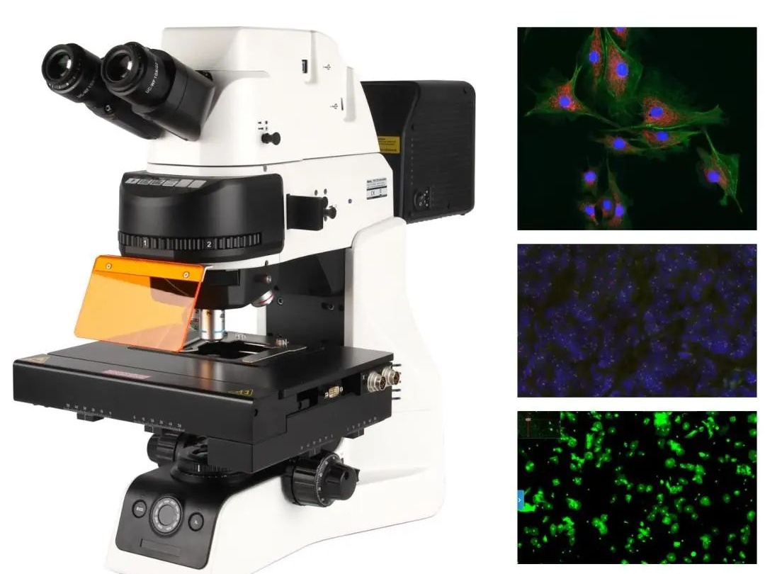
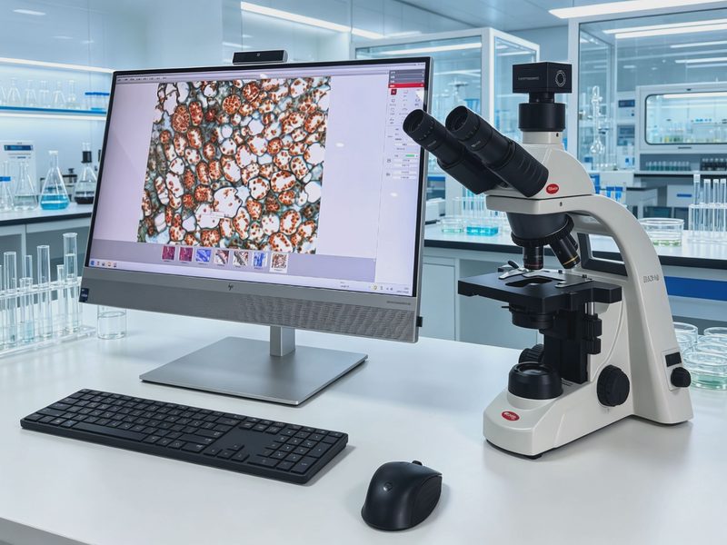
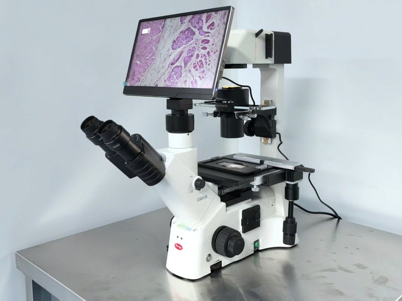
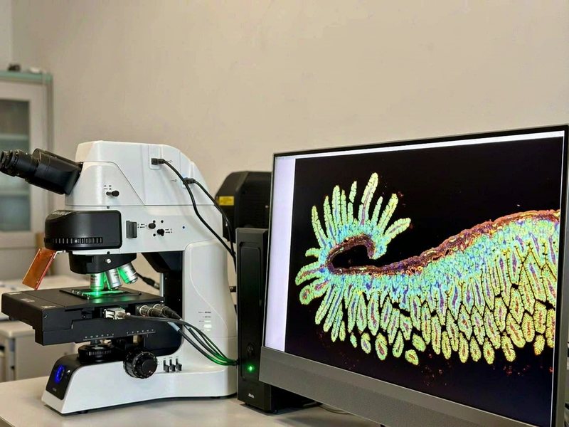
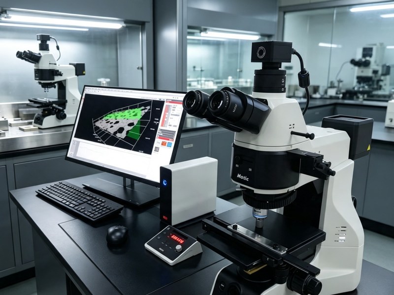
Delivered a PA53MET-BD-3D motorised metallurgical microscope with 3D surface topography analysis to the centre, for microstructural observation of diamond and CBN products, bond and interface analysis, coating thickness measurement and 3D surface assessment.
Superhard materials are a traditional strength of Henan's industry, with a dense cluster of diamond, cubic boron nitride and tooling manufacturers around Zhengzhou. Inspection in this field is hard for three reasons: the samples are extremely hard, the structures are very fine, and the acceptance criteria depend heavily on morphology.
1. What the centre needed to solve
The engineering technology centre does more than teaching. It provides technical services to local companies: particle morphology and size of diamond monocrystals and polycrystalline grades, microstructure and porosity of bonds (cobalt, resin, ceramic), interface bonding between diamond and matrix, thickness and uniformity of Ti or Ni coatings, and wear-surface morphology.
These used to be spread across several instruments: stereo microscopes for particle shape, ordinary metallurgical microscopes for structure and interface, manual image measurement for coating thickness, and verbal description for surface relief. The data never formed a system, and the images in reports were hard to reproduce.
2. Why a fully motorised configuration
Superhard products often have large observation areas — a matrix cross-section can be more than ten millimetres wide — while the details of interest are micrometre-scale interfaces.
A manual stage struggles here: moving field by field, stitching seams do not line up, and the same location cannot be found again next time.
The motorised configuration addresses all three. Stage movement is repeatable and coordinates are recorded, so multiple fields stitch into one image; motorised focus with autofocus keeps the image sharp even when the surface is slightly tilted; and motorised objective switching matches light intensity and aperture automatically instead of requiring readjustment every time.
For the centre, the practical benefit is that the same sample, measured by different people on different days, can be aligned image to image.
3. Brightfield, darkfield and 3D topography
Diamond and bond materials differ greatly in refractive index, so interfaces blur together in brightfield. Darkfield collects only scattered light, bringing particle outlines, microcracks and pore edges forward; reading the two together makes interface judgement far more reliable.
3D topography solves the problem of describing relief only in words. Through depth-of-field stacking, images from different focal planes combine into one fully sharp frame while height information is recorded, so groove depth on a worn surface, protrusion height of particles and fracture relief can be read directly. That is far more convincing than "it looks fairly flat".
4. Delivery and training
After arrival the system was installed, aligned and calibrated, with two training rounds for teachers and technicians: one on daily operation and image acquisition standards, one on 3D topography measurement and reporting.
Parameter templates for common inspection tasks — diamond particle morphology, matrix microstructure, coating cross-section, wear surface — were compiled into a handbook, so anyone taking over can produce consistent results by following the template.
5. Outcome
The instrument now serves both teaching and external testing. In teaching it supports microscopy experiments in materials forming and superhard products; in external service it provides microstructure and morphology analysis for local manufacturers, with reports including images and measurement data.
For an engineering technology centre, an instrument has to teach students and take commercial work at the same time. Stability, reproducibility and data output matter more than headline specifications.