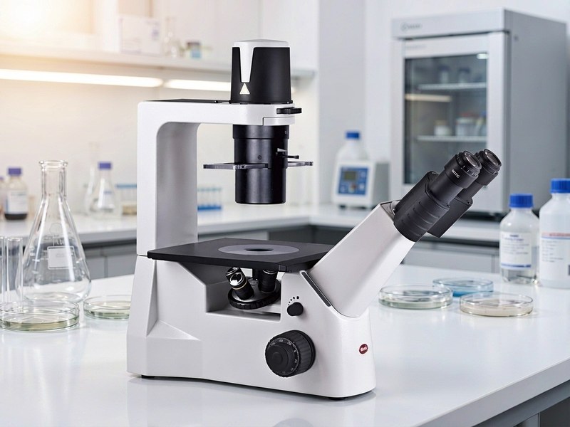
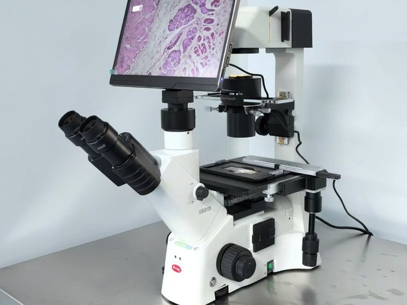
Phase Contrast Microscope Specifications
1. Optical system and objectives
• Infinity corrected optics with a parfocal distance of 45 mm or 60 mm, matched to the stand
• Phase objectives marked PH1, PH2 or PH3, each paired with one condenser annulus
• Phase objectives usually have 10 to 20 percent lower numerical aperture than brightfield versions, which is normal
• Common magnifications of 4X, 10X, 20X and 40X, with 10X and 20X most used for live cells
• Plan achromat or plan semi apochromat objectives with a field number of 22 mm or more
2. Annulus and phase plate matching: the key item
• Every phase objective needs its matching annulus, and annuli cannot be mixed across magnifications
• The condenser should be a phase type with a centring adjustment for the annulus
• The annulus ring diameter should be within 5 percent of the objective phase ring, or contrast drops and halo grows
• A turret with multiple annuli is better than single slide in plates, since no re centring is needed after changing magnification
• Note: a badly centred annulus is the most common cause of soft phase images and heavy halo
3. Contrast type and halo
• Positive phase renders cells darker than background and is the default for live cell work
• Negative phase renders cells brighter than background and suits some low contrast samples
• Halo is inherent to phase contrast and becomes stronger with thicker samples and higher numerical aperture
• High aperture phase objectives give stronger contrast but heavier halo, while low aperture types are softer
• Note: stopping down the aperture does not remove halo, it only costs resolution
4. Sample and vessel compatibility
• Best suited to live cells, unstained specimens and thin transparent samples, ideally 10 micrometres or less
• Plastic dishes and flasks have uneven, thick bases that weaken contrast, so glass bottom dishes are preferred
• Thick sections and tissue blocks are better observed with DIC or brightfield
• Slides and coverslips should match 0.17 mm thickness, since thicker coverslips add spherical aberration
• Both adherent and suspension cells work, with suspension samples allowed to settle or spun onto slides first
5. Condenser and illumination
• The phase condenser working distance must clear the dish height, with long working distance types for flasks
• LED or halogen illumination with adjustable, stable brightness, since phase is sensitive to uniformity
• An adjustable field diaphragm helps confine illumination and lift contrast
• A green filter improves resolution and contrast on colourless specimens
• Note: uneven illumination becomes visible banding in phase, so check field uniformity with a blank slide
6. Stage and focusing
• Mechanical stage with at least 75 by 50 mm travel, specimen holder and vernier scale
• Coaxial coarse and fine focus with a fine graduation of 2 micrometres or finer
• Long working distance models need stated clearance under the stage for flasks and multiwell plates
• Adapters for 96 well plates, flasks and dishes should be available
• Focus drift resistance matters for long term live cell observation
7. Digital imaging
• Trinocular head with a camera port splitting light 80 to 20 or 70 to 30
• Sensor of 5 megapixels or more, supporting long exposure and low gain settings
• Phase images have low contrast, so a bit depth of 10 bits or more avoids losing detail in compression
• Long term observation needs temperature and CO2 control together with focus drift compensation
• Note: phase images leave little room for post processing, so detail not captured cannot be recovered
8. Operating environment
• Ambient temperature from 15 to 30 degrees Celsius, away from vibration and direct sunlight
• Live cell work needs a heated stage or stage incubator holding 37 degrees Celsius within half a degree
• CO2 versions need a separate gas line and concentration control, normally 5 percent
• Objectives and condenser need dust protection, and the annulus turret should be checked for centring
9. Where procurement goes wrong
• Buying phase objectives alone without the matching annulus condenser, so no contrast forms at all
• Choosing a fixed, non centring annulus that cannot be aligned, leaving heavy halo that cannot be corrected
• Observing live cells in plastic dishes, where contrast stays poor and the equipment gets blamed