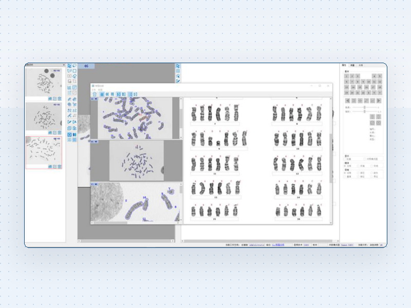
Chromosome Karyotyping System Specifications
1. Imaging system
• High resolution cooled camera from 5 megapixels, monochrome or colour
• Cooling of at least 10 degrees below ambient to lower noise in long exposure and weak staining
• Camera matched to the light path with a field number of 22 mm or more
• Bit depth of 12 or more to preserve banding detail
• Live preview and automatic exposure
2. Objectives and resolution: the key item
• 10X low power for whole slide scanning and metaphase search
• 100X oil immersion at 1.25 numerical aperture or better for band level analysis
• Resolution sufficient to resolve 400 bands, with 550 or more for high resolution work
• Plan apochromatic objectives to avoid chromatic fringes at chromosome edges
• Note: band resolution decides whether subtle structural abnormalities are detectable and is the core specification
3. Automatic scanning and metaphase detection
• Motorised stage scanning covering a standard slide
• Metaphase detection by staining intensity and morphology, located and marked automatically
• Whole slide scan at 10X within 3 minutes
• Detection accuracy must be verified on site, since debris and overlapping cells cause false positives
• Manual addition and skipping supported
4. Karyotype sorting and analysis
• Automatic pairing of chromosomes 1 to 22 plus sex chromosomes
• Manual correction by dragging, flipping, rotating, splitting and merging
• Banding methods G, R, C, Q and N matching the laboratory staining protocol
• Assisted marking of deletion, translocation, inversion and duplication
• Results reported under ISCN nomenclature
5. Database and case management
• Case database with patient data, specimen type, referring department and result
• Retrieval of historical cases and follow-up
• Multiple users with tiered rights for entry, review and final approval
• Original images and processing records retained together
• Backup and restore mechanism
6. Reporting
• Customisable templates for institution name, layout and terminology
• Output including the karyogram, original metaphase image and conclusion
• Export to PDF or Word and printing
• Electronic signature and three level review
• HIS and LIS integration with interface documentation
7. Compliance and interfaces
• Operation logs and audit trail
• Immutable data with read-only original images
• Conforming to clinical laboratory quality requirements
• Bidirectional connection with pathology and laboratory systems
8. Power and environment
• Power supply AC 220 V, 50 Hz
• Operating temperature 20 to 25 degrees Celsius
• Relative humidity up to 70 percent
• Anti-vibration table, since 100X oil work is vibration sensitive
• Darkened or controlled lighting
9. Where procurement goes wrong
• Only chromosome analysis system is specified without band resolution, and 300 to 400 bands arrive, missing subtle translocations
• Metaphase detection accuracy is poor, so slides are still found by eye and no efficiency is gained
• No HIS or LIS interface, so reports are retyped by hand with doubled workload and errors