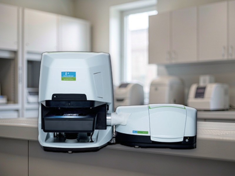
Infrared Microscope Specifications
1. Optical system
• Visible and infrared light paths, the visible for alignment and the infrared for acquisition
• Path change by motor or slider, without moving the sample
• Gold coated or moisture protected optics for humid environments
• Purgeable with dry air or nitrogen to suppress water vapour and CO2 interference
2. Spectral range and resolution: the key item
• Mid infrared range 4000 to 650 reciprocal centimetres as standard, extended models to 400
• Resolution of 4 reciprocal centimetres or better, with 1 to 2 needed for fine structure
• Wavenumber accuracy better than 0.1 reciprocal centimetres, verified on polystyrene
• Indicative signal-to-noise better than 10000 to 1 for a single point at 4 over one minute
• Note: infrared microscopy gives far less signal than a bench FT-IR, so signal-to-noise follows spot size
3. Detectors
• MCT detector, high sensitivity and fast, needing liquid nitrogen, the usual choice for microscopy
• DTGS detector, room temperature without nitrogen, lower sensitivity, suited to large spots
• Cooling method must be stated clearly, nitrogen dewar or electrically cooled, as it drives maintenance
• Nitrogen capacity and refill interval should be written into acceptance terms
4. Objectives and ATR
• Infrared objectives at 15X and 32X, both reflective Cassegrain, never ordinary glass optics
• ATR objectives in diamond, germanium or silicon for pressing onto opaque and thick samples
• Spot size down to 10 micrometres with a 32X ATR, to match the feature of interest
• Transmission mode needs KBr or ZnSe windows and samples under 10 micrometres thick
• Reflection mode suits coatings, films and surface contamination
5. Mapping stage
• Motorised X Y stage with at least 75 by 50 mm travel
• Minimum step of 1 micrometre or less for point by point mapping
• Chemical imaging from a grid of spectra, giving component distribution and peak intensity maps
• Line and area scans with predictable point counts and acquisition time
• Stage coordinated with visible imaging so the sample is seen before it is measured
6. Sample viewing and alignment
• Colour camera on the visible path for live viewing and point selection
• Crosshair or marker overlay so the measured point matches the visible location
• Spot position verified on a standard sample against the crosshair
• Large samples limited by stage travel and objective working distance
7. Software and libraries
• Acquisition with baseline correction, smoothing and normalisation
• Library search against commercial and user libraries, including mixtures
• Mapping processing with chemical imaging, principal component analysis and clustering
• Export to JCAMP-DX, CSV and TIFF
• Compliance features such as audit trail and electronic signature as options
8. Power and environment
• Power supply AC 220 V, 50 Hz
• Operating temperature 15 to 30 degrees Celsius, drift within 2 degrees per hour
• Relative humidity up to 60 percent, lower for KBr windows
• Away from vibration, since mapping is sensitive to it
• Safe storage and handling of liquid nitrogen must be arranged in advance
9. Where procurement goes wrong
• Only infrared microscope is specified without detector type or spot size, and a 100 micrometre spot arrives, making micro analysis impossible
• An MCT detector is supplied without a nitrogen routine, so it stays warm and loses sensitivity
• No ATR objective, so opaque and thick samples cannot be measured without destructive preparation