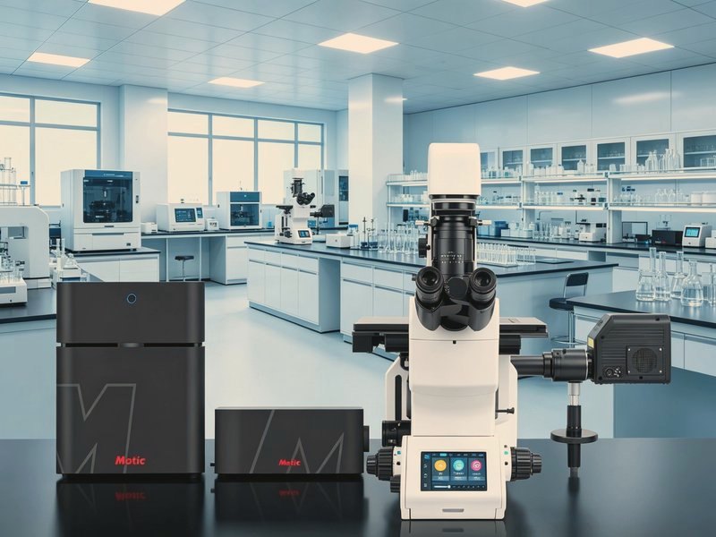
Laser Confocal Microscope Specifications
1. Optical and scanning system
• Infinity corrected optics with 45 or 60 mm parfocal distance
• Point scanning by galvo mirrors as standard, spinning disk for fast live cell work
• Scan head integrated with the stand or separate, the latter being easier to upgrade
• Light path supporting brightfield, fluorescence and confocal modes
2. Resolution and imaging performance
• Lateral resolution around 200 nm with a 60X 1.4 oil objective at 488 nm excitation
• Axial resolution around 500 to 700 nm
• Scan resolution up to 4096 by 4096 pixels
• A pinhole at 1 Airy unit balances resolution and light throughput, smaller means thinner slices and weaker signal
• Note: resolution follows objective numerical aperture and wavelength, and quoted figures come from the highest aperture oil objectives
3. Lasers and detection channels
• Solid state lasers, commonly 405, 488, 561 and 640 nm, at 10 to 50 mW output
• Laser power adjustable in software, with acousto-optic modulation or neutral density filters
• Three to four detection channels with photomultiplier or GaAsP detectors
• Spectral detection with up to 32 channels for unmixing overlapping fluorophores
• Simultaneous multichannel imaging requires attention to crosstalk and scan delay
4. Scan modes and speed
• Standard scanning at 512 by 512 giving 1 to 2 frames per second
• Bidirectional and resonant scanning raising this to 4 to 30 frames per second depending on model
• Zoom from 1 to 32 times, enlarging a region of interest without losing resolution
• Fast dynamics such as calcium imaging or blood flow need a resonant head or spinning disk
• Speed trades against signal-to-noise, and fast scanning usually needs more laser power or binning
5. Pinhole and optical sectioning
• Continuously adjustable pinhole from 0 to 1000 micrometres
• Pinhole set in Airy units, one unit equalling one optical section thickness
• Z step of 10 nm or less with a piezo stage, enabling Z stacks and 3D reconstruction
• Section thickness around 0.8 micrometres with a high aperture oil objective and above 5 at low power
• Z drift compensation or autofocus is essential for long live cell runs
6. Stage and live cell incubation
• Motorised X Y stage with at least 100 by 75 mm travel and 1 micrometre repeatability
• Accepting 35 mm dishes, multiwell plates, chamber slides and standard slides
• Incubation holding 37 degrees Celsius within 0.5 and CO2 at 5 percent within 0.2
• Multipoint time lapse across multiwell plates
• Objective working distance must still be respected once the chamber is fitted
7. Software and image analysis
• Multichannel acquisition, channel merging and false colour mapping
• 3D reconstruction by maximum intensity projection, orthogonal views and surface rendering
• Colocalisation analysis with Pearson and Manders coefficients
• Fluorescence intensity quantification, area and morphology measurement, particle counting
• Deconvolution and large area stitching
• Export to TIFF, OME-TIFF and AVI
8. Power and environment
• Power supply AC 220 V, 50 Hz, with a stabiliser or UPS recommended
• Operating temperature 20 to 25 degrees Celsius, drift within 1 degree per hour
• Relative humidity up to 70 percent, avoiding condensation
• Anti-vibration table, air or marble, away from lifts, compressors and corridors
• Darkened or controlled lighting so ambient light does not enter the detection path
9. Where procurement goes wrong
• Only confocal microscope is specified without laser wavelengths or channel count, and two channels arrive, making double labelling impossible
• No incubation chamber and no autofocus, so cells drift out of focus during time lapse and the data are lost
• The stage does not match the vessels in use, so a 96 well plate cannot be imaged well by well