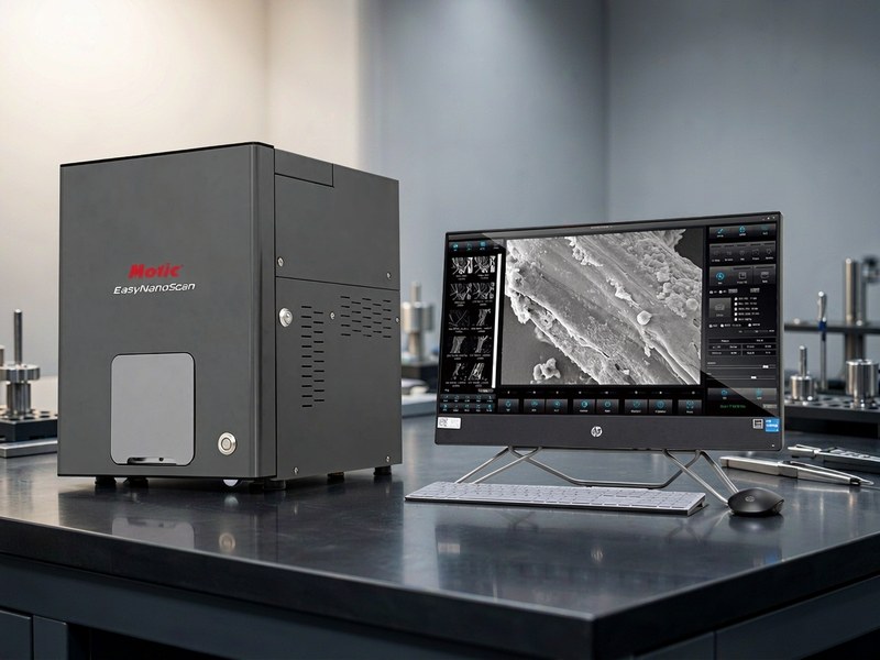
Benchtop SEM Specifications
1. Electron optics
• Tungsten filament or CeB6 single crystal source, tungsten being usual on benchtop models
• Short, compact column allowing the instrument to sit on an ordinary laboratory bench
• Magnetic lens focusing, with autofocus and automatic stigmation on some models
• No dedicated room required, though an independent earth is still needed on some models
2. Resolution and magnification: the key item
• Indicative resolution of 30 nm or better at 30 kV in secondary electron mode, depending on model and conditions
• High end benchtop models reach 10 to 15 nm
• Magnification 20X to 100000X digital, low power for overview and high power for surface detail
• Resolution depends on accelerating voltage, working distance and sample conductivity, and quoted figures come from ideal specimens
• Note: non-conductive samples need sputter coating or low vacuum mode, otherwise charging prevents imaging
3. Accelerating voltage
• Accelerating voltage adjustable from 5 to 15 kV, some models from 1 to 30 kV
• Low voltage from 1 to 5 kV for surface detail with less charging and beam damage
• High voltage above 15 kV penetrates deeper, suited to thicker or heavy element samples
• More voltage steps mean greater adaptability
4. Detectors
• Backscattered electron detector for compositional contrast, heavy elements bright and light dark, showing phase distribution
• Secondary electron detector for surface topography with strong depth cues
• Four-quadrant backscattered detector for mixed compositional and topographic imaging as an option
• Low vacuum mode on some models for non-conductive or moist samples
5. Chamber and stage
• Chamber taking stages of 25 to 50 mm diameter, several specimens at a time
• Manual X Y Z stage or three-axis motorised, travel around 30 to 50 mm
• Maximum specimen height limited by working distance, typically 30 mm or less
• Tilt and rotation as options, for fracture surfaces and side walls
6. Vacuum system
• Turbomolecular plus roughing pump, pump-down in 3 minutes or less
• Vacuum of 10 to the minus 3 pascal or better in high vacuum, 10 to 100 pascal in low vacuum
• Fast sample exchange without long venting cycles
• Periodic pump oil change or turbopump check, since poor vacuum degrades resolution directly
7. Imaging and elemental analysis
• Digital imaging with live display, capture, recording and measurement of distance, angle and area
• Export to TIFF, JPEG or BMP with the scale bar embedded
• Energy dispersive spectroscopy as an option for qualitative and quantitative elemental analysis, mapping and line scans
• EDS detectors liquid nitrogen cooled or electrically cooled, element range from beryllium to uranium depending on detector
• 3D surface reconstruction as an option, by tilt imaging and height calculation
8. Power and environment
• Power supply AC 220 V, 50 Hz, with a stabilised supply recommended
• Operating temperature 15 to 30 degrees Celsius, drift within 2 degrees per hour
• Relative humidity 40 to 70 percent, avoiding condensation
• Away from strong vibration and magnetic fields, not sharing a circuit with heavy equipment
• Independent earth with resistance of 4 ohm or less
9. Where procurement goes wrong
• Resolution of 10 nm is quoted without test conditions of voltage, working distance and specimen type, unreachable on biological or polymer samples
• No low vacuum mode and no coating equipment, so non-conductive samples simply cannot be imaged
• EDS is left out at purchase, making later retrofits expensive and impossible on some models