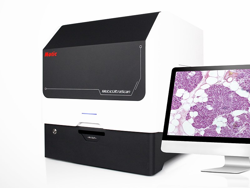
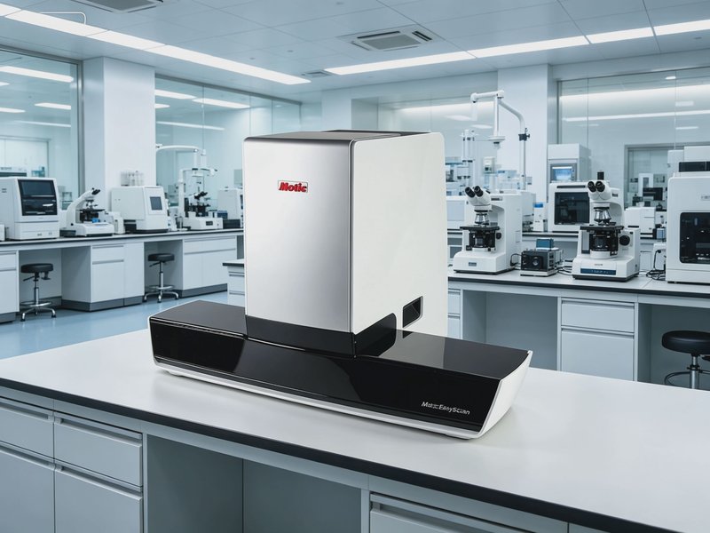
Whole Slide Scanner Specifications
1. Scanning principle and light path
• Brightfield transmitted scanning as standard, multicolour fluorescence optional
• Line scan with continuous motion, or area scan with step-and-stitch
• Autofocus by image contrast or laser assist, focusing per field in real time
• Automatic white balance and illumination uniformity correction, leaving no seams after stitching
• Z-stack scanning to pick the best focal plane in thick sections
2. Objectives and resolution: the key item
• 20X objective as standard, 40X optional
• Numerical aperture of 0.75 or more at 20X, 0.95 or more at 40X
• Indicative resolution of 0.25 micrometre per pixel at 20X and 0.12 at 40X
• Optical and pixel resolution stated separately, interpolation adds no real detail
• Note: pathology diagnosis generally needs 20X or higher, with 40X for nuclear detail
3. Speed and throughput
• A 15 x 15 mm tissue area scanned in 60 seconds or less at 20X brightfield
• Continuous loading of 6 slides or more, with 8 and 12 slide options
• Unattended batch scanning, moving to the next slide automatically
• Speed and image quality assessed together, since over-compression to gain speed loses diagnostic information
4. Slide loading and compatibility
• Standard slides of 25 x 75 mm, 1.0 to 1.2 mm thick
• Compatible with common mountants and stains such as HE, Pap and immunohistochemistry
• Automatic barcode reading with laboratory information system integration as an option
• Magazine removable as a unit for batch loading
5. Image output and formats
• Output in SVS, TIFF, JPEG2000 or a proprietary format, with a statement on exporting to common formats
• Pyramidal multi-resolution storage for fast zooming
• Export of single fields and of annotated regions
• Colour management following an ICC workflow for consistency between displays
6. AI analysis and viewing
• Optional AI modules for cell counting, nuclear-cytoplasmic ratio, immunohistochemistry quantification and chromosome analysis
• Viewing over the local network or in a browser, without a dedicated workstation if required
• Measurement, annotation, capture and sharing for consultation
• AI results traceable through model version, parameters and source image
7. Network and storage
• A 20X whole slide image runs from 200 MB to 1 GB, so size storage by daily volume
• Dedicated NAS or server recommended, archived by case
• Gigabit network as a minimum, 10 gigabit where several people view at once
• Regular backup, since images are primary medical or research data
8. Power and environment
• Power supply AC 220 V, 50 Hz
• Operating temperature 5 to 40 degrees Celsius, relative humidity up to 80 percent, dust kept out
• UPS recommended, an interrupted scan loses the current slide
• Clean the light path and stage guides periodically
9. Three points where procurement often goes wrong
• Only 40 megapixels is quoted without objective power or true resolution, and interpolated images cannot be used for diagnosis
• Speed is quoted on the smallest tissue area, while a full slide takes several times longer
• Output is a closed format with no export, which blocks later AI or consultation integration