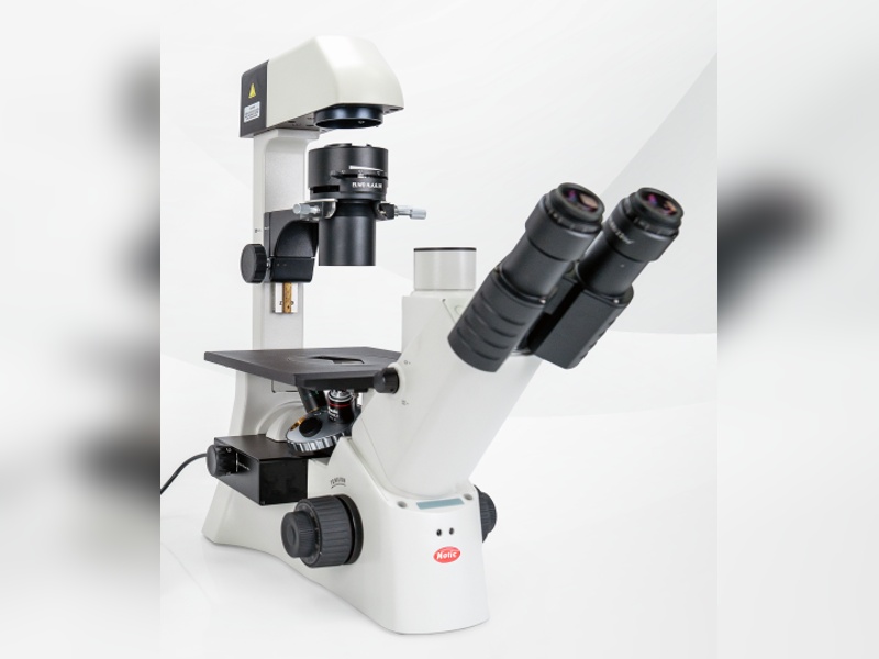
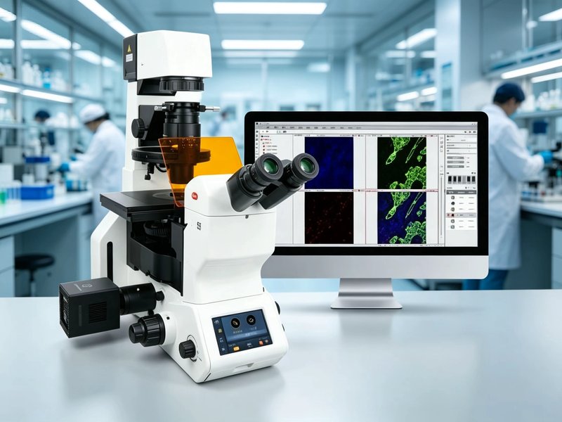
Fluorescence Microscope Specifications
1. Optical system
• Infinity-corrected optical system, incident fluorescence and transmitted brightfield sharing the same objectives
• 45 mm parfocal distance
• Fluorescence and brightfield paths switched by turret or slider without refocusing
• Stray light suppressed in the light path for a dark background and high signal-to-noise ratio
2. Fluorescence path and channels: the core item
• Each channel is built from an excitation filter, a dichroic mirror and an emission filter
• Blue channel B, excitation 460 to 495 nm and emission 510 to 550 nm, for FITC, GFP and Calcein
• Green channel G, excitation 510 to 560 nm and emission 575 to 625 nm, for TRITC, Rhodamine and DsRed
• Ultraviolet channel U, excitation 340 to 390 nm and emission 420 to 460 nm, for DAPI and Hoechst
• Channel count and combination selected around the labels used, commonly two, three or four
• Turret switching of 1 second or less, supporting sequential multicolour capture
3. Objectives
• Plan achromat or semi-apochromat objectives for fluorescence: 4X, 10X, 20X, 40X, 100X oil
• Indicative numerical aperture: 10X 0.30 or more, 20X 0.50 or more, 40X 0.75 or more, 100X 1.30 or more
• Higher numerical aperture collects fluorescence more efficiently, which shows clearly on weak samples
• Objectives matched to 0.17 mm coverslips, oil objectives using fluorescence grade immersion oil
4. Light source
• LED source, switchable per channel, dimmable, no warm-up, life of 20000 hours or more
• 100 W mercury lamp, bright at specific lines but needing warm-up, short-lived and containing mercury
• Source stability affects quantitative fluorescence directly, LED is preferable for long sessions
• Some models offer stepped intensity at 25, 50 and 100 percent to reduce photobleaching
5. Head and eyepieces
• Trinocular head, 30 degree inclination, camera port available
• Split ratios typically 100:0 or 20:80, with 0:100 preferred for imaging
• 10X wide-field eyepieces, field number 20 mm or larger
• Eyepiece barrier filters available as an option for eye protection
6. Stage and focusing
• Mechanical stage, travel of at least 75 x 50 mm
• Coaxial coarse and fine focusing, fine step of 0.002 mm or less
• Heated stage and CO2 enclosure recommended for long-term live-cell imaging
• Multi-well plate adapter available
7. Digital imaging: sensitivity is the key
• For fluorescence look at quantum efficiency and noise rather than pixel count
• Sensor size of 2/3 inch or larger, 5 to 12 megapixels, pixel size no smaller than 3 micrometres
• Cooled cameras, typically to minus 10 degrees Celsius, reduce dark current on weak signals
• Exposure, gain and binning adjustable, with multichannel overlay and pseudocolour
8. Power and environment
• Power supply AC 220 V, 50 Hz
• A darkened room improves the result, ambient light raises the background
• Operating temperature 5 to 40 degrees Celsius, relative humidity up to 80 percent
• Store labelled samples in the dark to slow label decay
9. Three points where procurement often goes wrong
• Only fluorescence microscope is specified with no channel list, and a single B channel arrives, making double labelling impossible
• Cross-talk between channels is not assessed, so multicolour samples bleed into each other and the data are unusable
• The camera has a high pixel count but a small sensor and no cooling, so noise overwhelms weak fluorescence