Project Overview
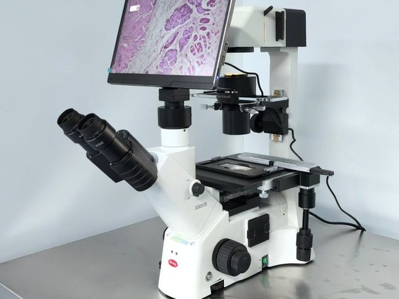
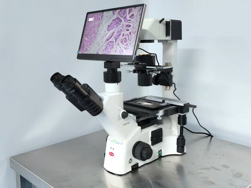
Delivered an AE31E inverted biological microscope with digital imaging to Henan Cancer Hospital, for morphological observation and density assessment of cytology specimens and cultured tumour cells, supporting routine testing, research work and case review.
In a cancer hospital, several of the samples seen every day resist being pressed under a coverslip: cytology smears from pleural and ascitic fluid or fine-needle aspiration, tumour cell lines under culture and drug-sensitivity work, and increasingly organoids and 3D cell spheroids. What they share is that they cannot be flattened, and should not always be stained and mounted. The ideal approach is to observe them directly in the culture vessel.
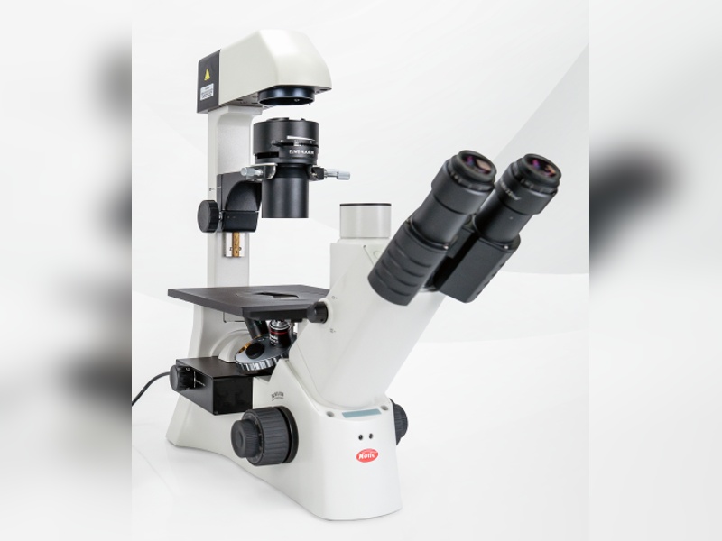
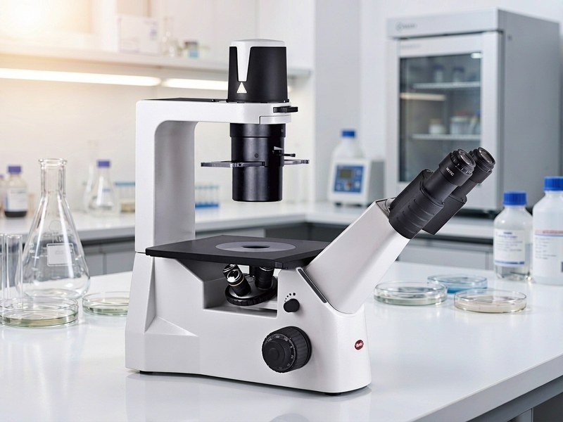
An inverted microscope is built for exactly this. Objectives sit below and samples are placed from above, so dishes and flasks go straight onto the stage and cell morphology, attachment and density can be read through the base. Long working distance objectives are essential here — thick vessel walls and deep liquid layers put the focal plane out of reach for ordinary optics.
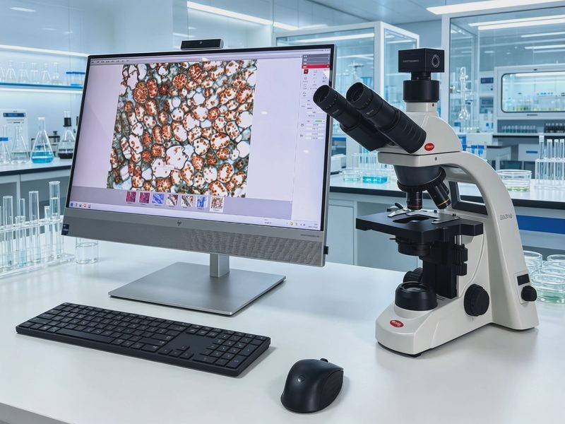
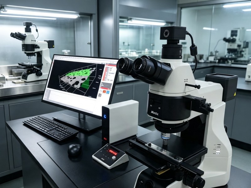
The hospital received an AE31E inverted biological microscope with digital imaging. Installation, commissioning and optical alignment were completed, and laboratory and research staff were trained on brightfield observation, focusing and stage movement, image capture and exporting images for reports. The system is now part of routine testing and research workflows, with captured images used directly in case review, experimental records and archiving.