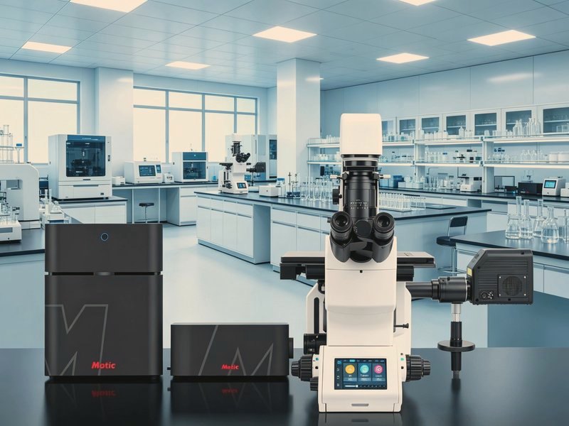
Confocal Microscopy: Why It Can Cut a Cell into Thin Optical Slices
When imaging thicker samples — cell clusters, tissue sections, material cross-sections — a conventional fluorescence microscope collects light from above and below the focal plane as well, blurring the image. Confocal microscopy solves exactly this problem by optically sectioning a thick sample so it can be viewed layer by layer.
1. The core principle: one point in, one plane out
The key lies in the word "confocal": the point light source, the focal point on the sample and the pinhole in front of the detector are arranged in mutually conjugate positions. A laser is focused by the objective onto a single point in the sample. Of the fluorescence emitted from that point, only light that can pass through the pinhole reaches the detector; stray light from above and below the focal plane is blocked. Only information from the focal plane is recorded — an "optical section".
To build a two-dimensional image, the system scans point by point: galvanometer mirrors move the light spot across the sample, collecting point by point and building up line by line. Combined with stepwise Z movement, this yields a stack of optical sections that software reconstructs into a three-dimensional structure.
2. What it does better than a conventional fluorescence microscope
• Optical sectioning — defocus blur is removed, so thick samples yield clear cross-sections;
• Better contrast — background stray light is suppressed and fine structure stands out;
• 3D reconstruction — stacked Z-sections produce a three-dimensional result;
• Quantification — fluorescence intensity is measurable, supporting co-localisation and quantitative analysis.
The trade-offs matter too: laser sources are costly, scanning is relatively slow, and phototoxicity and photobleaching are stronger — a real consideration for live-cell work.
3. Key components and selection
• Lasers — common wavelengths such as 405/488/561/640 nm, chosen to match the fluorescent probes;
• Scanning method — galvo scanning is standard, while spinning-disk confocal is faster and better suited to live cells;
• Detectors — photomultiplier tubes (PMT) are general-purpose, while spectral detectors can split channels;
• Pinhole size — a small pinhole gives higher resolution but weaker signal; a large one gives stronger signal but poorer sectioning.
4. What confocal microscopy can do
• Cell biology — organelles, cytoskeleton and membrane protein localisation;
• Live-cell imaging — calcium signals, membrane transport, cell migration;
• Multiplex fluorescence — simultaneous imaging of multiple labels and co-localisation analysis;
• Materials research — coating cross-sections, luminescent materials, semiconductor structures.
Conclusion
Confocal microscopy turns "thick samples you cannot see clearly" into "layers you can see one by one" — a key step in fluorescence imaging's move from seeing the whole to seeing the structure. Before choosing a system, be clear about one thing: are you imaging thin, fixed samples, or thick, living ones? The answer changes the configuration completely.