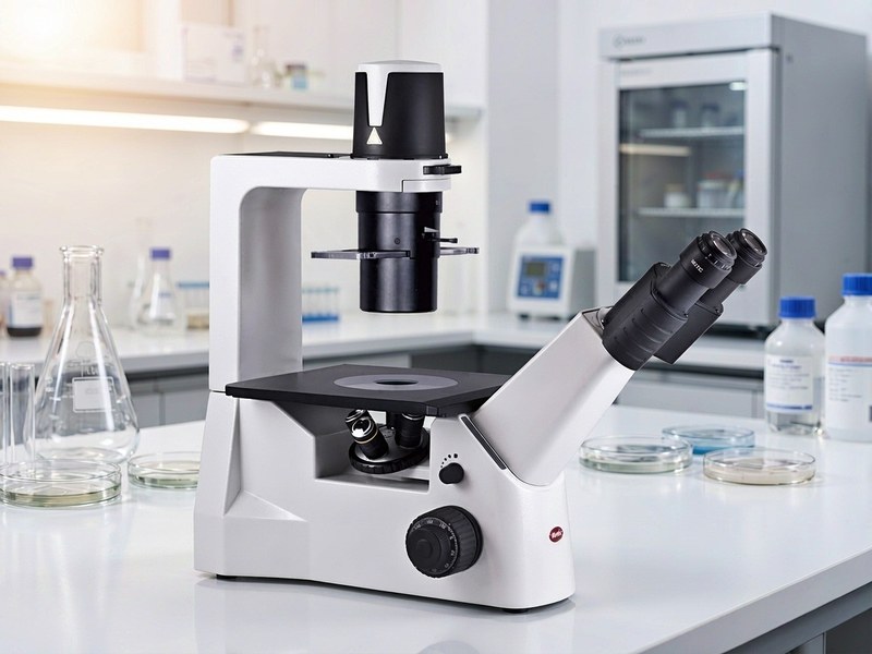
The Inverted Microscope: Why Live-Cell Work Depends on It
In cell culture, patch clamp work and micromanipulation, the instrument used most often is not the familiar upright microscope but the inverted one. Its objectives sit below the stage and look upward, and that apparently simple change of layout solves a whole set of practical problems in live-cell observation.
1. The difference from an upright microscope
On an upright microscope the objective is above and light passes up through the specimen; on an inverted microscope the positions are swapped, with the objective below and the light source above. The sample therefore sits on a stage above the objective, and it can be a culture dish, a flask or a multi-well plate: the vessel is never opened or turned over, and the cells can be observed directly. That is the precondition for any live-cell experiment.
2. Why live-cell work depends on it
Living cells have to be kept in a dish, in culture medium, at a controlled temperature and gas atmosphere. The inverted design leaves ample space for large vessels and at the same time allows the same site to be observed repeatedly without disturbing the culture, recording growth, division and migration over time. Switch to an upright microscope and either the specimen has to be very thin or the vessel will not fit at all; the experiment simply cannot be done.
3. Typical applications
Culture and morphological observation is the most basic application. Patch clamp, microinjection and other electrophysiology and micromanipulation work are almost all carried out on an inverted microscope platform, because microelectrodes and manipulators need free access from above. Add IVF, stem cell research and organoid culture. As a rule, any experiment that keeps cells in a vessel and needs to see them alive is run on an inverted microscope.
4. What to look for when choosing one
The first point is long working distance objectives: ordinary objectives hit the base of the dish, so inverted work needs long, sometimes extra-long, working distance optics. The second is the observation mode: brightfield for routine morphology, phase contrast or DIC for cells with little contrast, and a fluorescence light path where labels are involved. The third is environmental control: long-term live imaging needs a heated stage, a CO2 enclosure or even an integrated incubator. The fourth is the interface for expansion, so that manipulators, cameras and perfusion systems attach easily.
5. The platform trend
Modern inverted microscopes behave more like an experimental platform than a viewing instrument. They have to accept a wide range of accessories, image stably over long periods, and offer enough mechanical precision and expandability to work with patch clamp rigs and live-cell workstations. When choosing, think about the experiments you may be running two or three years from now and leave room to grow.
Put simply, the value of an inverted microscope is not in how much it magnifies but in making live-cell observation feasible, controllable and repeatable. Get this platform right before starting cell or electrophysiology work, and everything downstream goes more smoothly.